




Medical education
Clinical skills
Volume 205 - Issue 4
Pitting and non-pitting oedema
Authors: Elizabeth Whiting and Madeline E McCready
Med J Aust 2016; 205 (4): 157-158. || doi: 10.5694/mja16.00416
Published online: 15 August 2016
Published online: 15 August 2016
The distinction is essential to determine aetiology and treatment
The distinction is essential to determine aetiology and treatment
Oedema can be divided into two types: pitting and non-pitting. These types are relatively easy to distinguish clinically and the distinction is essential to determine aetiology and treatment.
Oedema is the swelling of soft tissue due to fluid accumulation. Pitting is demonstrated when pressure is applied to the oedematous area and an indentation remains in the soft tissue after the pressure is removed (Box 1 and Box 2). Moreover, mild pitting oedema is best identified by applying pressure over an area of bony prominence. Non-pitting oedema refers to the lack of persistent indentation in the oedematous soft tissue when pressure is removed.1
In addition to the differentiation of pitting and non-pitting oedema, the pattern of distribution is reflective of the underlying aetiology. With pitting oedema, there may be bilateral dependent oedema of the lower limbs, generalised oedema or localised oedema. Non-pitting oedema generally affects an isolated area, such as a limb. There are two ways of describing the severity of pitting oedema. Most commonly, in the setting of peripheral oedema, severity is graded by its proximal extent, so that oedema located above the knee is more severe than oedema presenting below the knee. The alternative approach is based on depth and duration of pitting after the release of pressure (Box 3).2
A number of factors3 can be considered to be major contributors to the development of oedema:
increased intravascular hydrostatic pressure;
reduced intravascular oncotic pressure;
increased blood vessel wall permeability;
obstructed fluid clearance in the lymphatic system; and
increased tissue oncotic pressure.
The underlying pathophysiology for oedema explains the reason for pitting and non-pitting. Oedema is the accumulation of fluid in the interstitium. In normal circumstances, there is a balance between fluid leaking from capillaries and drainage by the lymphatics.3 In the setting of increased intravascular hydrostatic pressure, reduced oncotic pressure or where there is increased vessel wall permeability, fluid leaks out of vessels into the interstitial space. When external pressure is applied, extracellular fluid is displaced with increased drainage through the lymphatic system, creating an indentation that is visible in the skin and is described as pitting. When pressure is removed, the fluid slowly returns and the indentation disappears (see the video at mja.com.au). Lymphoedema, which is the most common form of non-pitting oedema, occurs when fluid accumulates in the interstitial space as a result of a reduction in lymphatic drainage. The application of pressure does not result in an indentation as there is an inability to drain fluid through the damaged lymphatic system.4 An uncommon form of non-pitting oedema, myxoedema, can occur as a result of accumulation of hydrophilic molecules in the subcutaneous tissue.5
The differentiation between pitting and non-pitting oedema, in addition to the pattern of distribution, reflects different pathophysiology and may therefore be helpful in identifying the underlying aetiology. Pitting peripheral lower limb oedema resulting from raised hydrostatic pressure can occur in congestive cardiac failure, venous insufficiency and as a result of the use of a calcium channel blocker. Generalised oedema may be seen in kidney disease, where reduced intravascular oncotic pressure occurs through protein loss or where increased vascular volume occurs through sodium and fluid retention. Localised pitting oedema is likely related to a local inflammatory process resulting in increased vessel wall permeability. Non-pitting oedema of an isolated area is consistent with failure of lymphatic drainage, which may rarely have a primary (hypoplastic) aetiology or, more commonly, a secondary (obstructive) aetiology. Secondary causes include external compression from a tumour, involvement of lymph nodes in metastatic disease, and lymph vessel damage following radiotherapy or following lymph node resection. Myxoedema is associated with thyroid disease.5
The underlying aetiology will guide treatment options. In the setting of congestive cardiac failure, treatment would generally include fluid restriction and diuretics (including spironolactone). Compression bandaging and leg elevation are useful for oedema related to venous insufficiency. In the setting of oedema related to inflammation, a general approach would include applying ice and elevation in addition to treating the underlying cause of the inflammatory process. Lymphoedema may be detected using a perometer and managed with compression garments and manual lymph drainage. Myxoedema may resolve with treatment of the underlying thyroid condition.
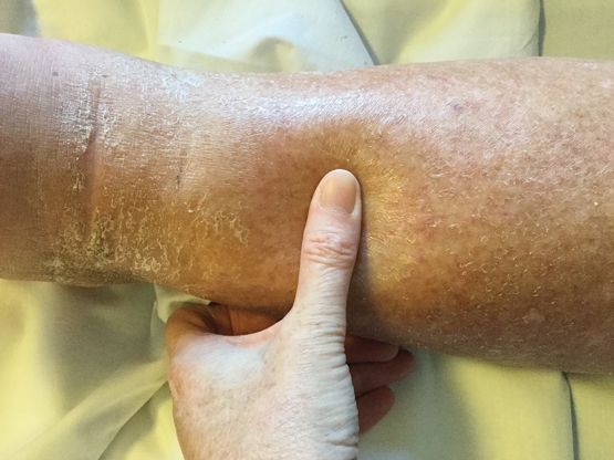
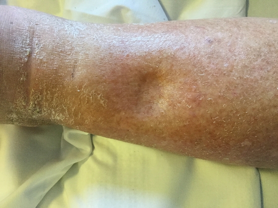
Box 3 – Alternative approach for grading the severity of pitting oedema based on depth and duration of pitting after release of pressure
Severity |
Description |
||||||||||||||
Grade 1+ |
A pit of up to 2 mm that disappears immediately |
||||||||||||||
Grade 2+ |
A pit of 2–4 mm that disappears in 10–15 seconds |
||||||||||||||
Grade 3+ |
A pit of 4–6 mm that may last more than 1 minute |
||||||||||||||
Grade 4+ |
A deep pit greater than 6 mm that may last as long as 2–5 minutes |
||||||||||||||
Adapted from Guelph General Hospital Congestive Heart Failure Pathway.2 | |||||||||||||||
Competing interests
No relevant disclosures.
References
- Talley NJ, O’Connor S. Clinical examination: a systematic guide to physical diagnosis. 6th ed. Sydney: Elsevier, 2010.
- Grey Bruce Health Network. Assessment of pitting edema. Ontario: Guelph General Hospital Congestive Heart Failure Pathway, 2009. http://www.gbhn.ca/ebc/documents/ASSESSMENTOFPITTINGEDEMA.pdf (accessed Apr 2016).
- Scallan J, Huxley VH, Korthuis RJ. Pathophysiology of edema formation. Capillary fluid exchange: regulation, functions, and pathology. San Rafael: Morgan and Claypool Life Sciences, 2010: pp 47-62. http://www.ncbi.nlm.nih.gov/books/NBK53445/ (accessed Apr 2016).
- Wiig H, Swartz MA. Interstitial fluid and lymph formation and transport: physiological regulation and roles in inflammation and cancer. Physiol Rev 2012; 92: 1005-1060.
- Trayes KP, Studdiford JS, Pickle S, Tully A. Edema: diagnosis and management. Am Fam Physician 2013; 88: 102-110.
Provenance: Commissioned; externally peer reviewed.
Fatty Liver Disease in Australia: A Narrative Review on the Epidemiology, Natural History, Prognostication and Management in People With Metabolic Dysfunction
Karl Vaz, Daniel Clayton-Chubb, William W. Kemp, Stuart K. Roberts, Ammar Majeed
Sex Differences in Management and Outcomes of People With ST-Elevation Myocardial Infarction, New South Wales, 2011–2020: A Retrospective Cohort Study
Adam C. Livori, Jedidiah I. Morton, Dianna J. Magliano, Jonathan E. Shaw
Rethinking Rheumatic Fever Diagnosis: Progress, Pitfalls and the Path Towards a Definitive Test
Anna P. Ralph, Rachel Webb, Tom Parks, Nicole J. Moreland, Asha C. Bowen, Benjamin Jones, Emma Ndagire, Andrea Z. Beaton
Sex Differences in Management and Outcomes of People With ST-Elevation Myocardial Infarction, New South Wales, 2011–2020: A Retrospective Cohort Study
Samia Kazi, Simone Marschner, Clara K. Chow
Distribution of Cardiovascular Disease Risk Based on the Updated 2023 Guideline-Recommended Australian Cardiovascular Disease Risk Algorithm and Comparison With the 2012 Algorithm: An Observational Study
Nina Lazarevic, Meghana Bhat, Grace Joshy, Danielle C. Butler, Mark Woodward, Anushka Patel, Rod T. Jackson, Garry Jennings, Rosemary Wyber, Ellie Paige, Emily Banks
They Brought the Hospital to Me
Hugh N. Burrill