Connect
MJA
MJA

Intensive rehabilitation in a patient with inclusion body myositis

Anupam Datta Gupta and Nigel Quadros
Med J Aust 2009; 190 (4): 208-209. || doi: 10.5694/j.1326-5377.2009.tb02352.x
Published online: 16 February 2009

Clinical record

In November 2006, a 59-year-old man was admitted to hospital with a 5-week history of dyspnoea secondary to type I respiratory failure, generalised weakness, poor mobility, and bilateral shoulder and knee pain. He had been diagnosed with inclusion body myositis in 1998. Comorbidities included systemic lupus erythematosus, pulmonary hypertension, interstitial pulmonary fibrosis (Figure, A), type 2 diabetes, obstructive sleep apnoea and gout; and he had undergone a bilateral total hip replacement, which enabled him to walk a limited distance unaided. Six months earlier, his general practitioner had noted a functional decline caused by a combination of poor pulmonary function, myositis and inactivity, which resulted in the patient requiring a wheelchair for mobility and help from his son with some activities of daily living. Methotrexate and azathioprine, which had been prescribed for management of the underlying connective tissue disease, were thought to be contributing to the respiratory failure and were withdrawn. Prednisolone therapy (50 mg daily) was begun, and some improvement in respiratory function was noted after 2 weeks. However, the patient did not regain baseline function and remained bed-bound, requiring assistance with all activities of daily living. Admission to a high-level residential care facility was considered, but the patient wanted to return home with his son as carer.

A rehabilitation physician was consulted 3 weeks after admission, with a view to improving the patient’s functional status so that he could return home. The patient was left-hand dominant with bilateral poor grip due to distal muscle weakness and metacarpophalangeal and interphalangeal joint contracture secondary to systemic lupus erythematosus. Muscle strength surrounding the shoulder girdles, hips and quadriceps declined from 4/5 to 2/5 bilaterally over a period of 6 months owing to myositis and inactivity. Advanced osteoarthritic changes were evident in the glenohumeral joints, resulting in pain (greater on the left side) and global restriction of shoulder movements. Moreover, an x-ray showed avascular necrosis of the head of the left humerus (Figure, B). X-rays of the knee joints and lower legs showed bilateral recurvatum deformity (hyperextension) and gross mediolateral instability, caused by quadriceps weakness. He could transfer from bed to chair and stand with the aid of a mechanical lifting device and moderate assistance from two people. Knee pain prevented him from standing unaided for more than 2 minutes.

An inpatient rehabilitation program was implemented by a multidisciplinary team comprising a rehabilitation physician, physiotherapist, occupational therapist and rehabilitation nurses. The program included strengthening exercises for weaker muscle groups, as well as training focused on building endurance to improve transfers and activities such as grooming, bathing and toileting. The patient was prescribed continuous home oxygen therapy (4 L/min) because of poor spirometry results (forced expiratory volume in one second [FEV1], ratio of FEV1 to forced vital capacity, and carbon monoxide diffusion in the lung were 60%, 91% and 21% of predicted values, respectively, with no bronchodilator response) and abnormal blood gas concentrations (Po2, 60 mmHg; Pco2, 26 mmHg; pH, 7.52). The myositis was monitored weekly by measuring erythrocyte sedimentation rate and levels of C-reactive protein and creatine kinase.

The patient underwent a left suprascapular nerve block to alleviate the left shoulder pain. The degrees of pain and disability were assessed before and after the procedure using the Shoulder Pain and Disability Index;1 the patient reported a 75% reduction of pain in the left shoulder and 25% improvement in shoulder disability. A carbon-fibre hinged orthosis was prescribed for each knee to correct the recurvatum deformity and improve gait (Figure, C). The orthoses controlled hyperextension during walking and corrected the instability of the knees. C-reactive protein and creatine kinase levels remained within the reference ranges over a period of 6 weeks.

After 6 weeks of rehabilitation, the patient required less oxygen (2 L/min) and maintained an oxygen saturation greater than 90%, with quick recovery after activity, indicating improved cardiovascular endurance. Also, his maximum heart rate 5 minutes after exercise decreased from 130 beats/min to 84 beats/min. The reduction in pain and dyspnoea resulted in improved transfers and ability to perform activities of daily living. In December 2006, after the 6-week rehabilitation period, he was able to walk 30 m with the aid of the orthoses and a walking frame, and he was discharged. Home-based exercises were implemented by his son, the nerve block was repeated every 4 months, and he continued to live at home until June 2008. In July 2008, he died due to pneumonia and septic shock.

This case highlights the capacity of therapeutic exercise, pulmonary rehabilitation, pain management and use of orthoses to reduce the impact of disability, restore function and potentially allow patients to live at home with family and community support. Our patient had several comorbid conditions that required specific management, in addition to general deconditioning after his acute illness.

Inclusion body myositis. This late-onset inflammatory muscle disease results in impaired muscle function, muscle atrophy and weakness, affecting both proximal and distal muscles. It accounts for 17%–30% of idiopathic inflammatory myopathies and can be associated with autoimmune diseases.2-4 In the past, patients with myositis were discouraged from exercising owing to a fear of increased muscle inflammation. However, studies in the 1990s reported that exercise might have a non-specific benefit.5 Patients with inflammatory muscle disease benefit from mild to moderate muscle training and endurance exercise, and muscle inflammation does not increase after exercise.6-8 A specifically tailored rehabilitation program improved our patient’s physical function without evidence of increased muscle damage.

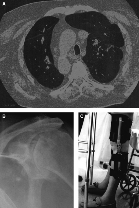
Interstitial pulmonary fibrosis. This group of lung diseases affects the interstitium of the lungs, eventually causing restrictive lung disease. Pulmonary rehabilitation — involving strength and endurance training for arm and leg muscles (eg, walking, cycling, lifting small weights), education on energy conservation and anxiety management, chest physiotherapy, and breathing techniques such as pursed lip and diaphragmatic breathing — has been shown to reduce dyspnoea, improve exercise capacity, enhance quality of life and reduce hospitalisation.9

Chronic shoulder pain from arthritis. This type of pain can be safely and effectively treated by suprascapular nerve block, which avoids the side effects of oral analgesics.10 In our patient, it was an essential component of the rehabilitation program because it enabled him to perform activities of daily living and use a walking frame. Intra-articular steroid injection was not used as it might have worsened the avascular necrosis of the head of the left humerus.

Genu recurvatum. This is an angular deformity (hyperextension at the knee) in the sagittal plane caused by quadriceps weakness. Mediolateral (valgus and varus) instability occurs in the coronal plane. Both deformities require three-point stabilisation. In our patient, orthoses with a free-motion joint and a hyperextension block controlled both deformities and allowed knee movement during walking.11 Gait training with such orthoses includes static weight shift and dynamic balancing exercise in parallel bars, followed by progression to a normal gait pattern.

Few residential care facilities in Australia specifically cater for disabled patients younger than 65 years. Our patient, who had an able and willing carer available, was highly motivated to return home. We believe that the option of active rehabilitation should always be explored, regardless of the apparent severity of underlying medical conditions.

  • Anupam Datta Gupta1
  • Nigel Quadros2

  • Department of Rehabilitation Medicine, Queen Elizabeth Hospital, Adelaide, SA.


Correspondence: anupamduttagupta@yahoo.com

Acknowledgements: 

We acknowledge Maree Braithwaite (Occupational Therapist), Nicolle Datson (Physiotherapist) and Jessica Quayle (Orthotist) of St Margaret’s Rehabilitation Hospital for assisting in the management of our patient. We also thank Dr Roger Hunt and Dr Alistar Bonin of Western Adelaide Palliative Care for reviewing the manuscript.

Competing interests:

None identified.

  • 1. MacDermid JC, Solomon P, Prkachin K. The Shoulder Pain and Disability Index demonstrates factor, construct and longitudinal validity. BMC Musculoskelet Disord 2006; 7: 12.
  • 2. Carpenter S, Karpati G, Heller I, Eisen A. Inclusion body myositis: a distinct variety of idiopathic myopathy. Neurology 1978; 28: 8-17.
  • 3. Lotz BP, Engel AG, Nishimo H, et al. Inclusion body myositis. Observations in 40 patients. Brain 1989; 112: 727-747.
  • 4. Rugiero M, Koffman B, Dalakas MC. Association of inclusion body myositis with autoimmune and autoantibodies. Ann Neurol 1995; 38: 333.
  • 5. Von der Kooi EL, Lindeman E, Riphagen I. Strength training and aerobic exercise training for muscle disease. Cochrane Database Syst Rev 2005; (1): CD003907.
  • 6. Alexanderson H, Lundberg IE. The role of exercise in the rehabilitation of idiopathic inflammatory myopathies. Curr Opin Rheumatol 2005; 17: 164-171.
  • 7. Varju C, Petho E, Kutas R, Czirjak L. The effect of physical exercise following acute disease exacerbation in patients with dermato/polymyositis. Clin Rehabil 2003; 17: 83-87.
  • 8. Arnardottir S, Alexanderson H, Lundberg H, Borg K. Sporadic inclusion body myositis: pilot study on the effects of a home exercise program on muscle function, histopathology and inflammatory reaction. J Rehabil Med 2003; 35: 31-35.
  • 9. Abramson MJ, Crockett PA, McDonald CF. COPDX: an update of guidelines for the management of chronic obstructive pulmonary disease with a review of recent evidence. Med J Aust 2006; 184: 342-345.
  • 10. Shanahan EM, Ahern M, Smith M, et al. Suprascapular nerve block (using bupivacaine and methylprednisolone acetate) in chronic shoulder pain. Ann Rheum Dis 2003; 62: 400-406.
  • 11. Datta Gupta A, Mahalanabis D. Genu recurvatum in hemophilia: a case report. Arch Phys Med Rehabil 2007; 88: 791-793.

Author

remove_circle_outline Delete Author
add_circle_outline Add Author

Comment
Do you have any competing interests to declare? *

I/we agree to assign copyright to the Medical Journal of Australia and agree to the Conditions of publication *
I/we agree to the Terms of use of the Medical Journal of Australia *
Email me when people comment on this article

Online responses are no longer available. Please refer to our instructions for authors page for more information.