


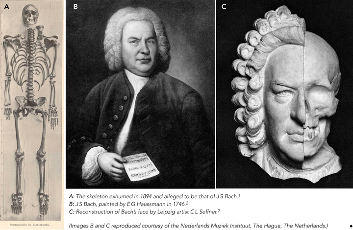
There has been much conjecture about the authenticity of this skeleton and it has been examined several times to establish its identity. In 1894, anatomist Wilhelm His reconstructed the face1,2 and, with remarkable foresight, took into account the possibility of future research, discussing the possibility of “our descendants in later centuries wanting to examine the skull”.2 In 1949, surgeon Wolfgang Rosenthal noted abnormalities of the skeleton that corresponded with those he saw in radiographs of living church organists, leading him to propose a musculoskeletal condition, Organistenkrankheit (“organist’s disease”).3
Several years after Bach’s burial in July 1750, the exact location of his grave was unclear.1-6 Based on oral tradition, it was in the graveyard surrounding St Johanniskirche in Leipzig, “six paces away from the south portal”.4 This oral tradition apparently originated in 1894 from a 75-year-old man, who in turn was informed about the location 60 years earlier by a 90-year-old gardener employed at the graveyard.4 Composer Robert Schumann attempted unsuccessfully to find Bach’s grave (in 1836), as did Leipzig historians Carl Gretschel and Heinrich Heinlein, who both published books on the history of the graveyard (in 1836 and 1844, respectively).4
Before the 1894 exhumation, Leipzig’s director of archives Gustav Wustmann determined that Bach was buried in one of the so-called superficial graves, whose location was not registered.4 Compounding the difficulty, in the year of Bach’s death, no graves were marked with a memorial stone. However, unusually for the time, Bach was buried in an oaken coffin (of the 1400 people who died in Leipzig in 1750, only 12 were buried in such a coffin).4
Thus, when — according to local oral tradition — an oaken coffin containing the skeleton of a man aged about 65 years was found at a depth of 2.37 m in the specified area of the graveyard on 22 October 1894, it was, after the research by His, believed likely to be the remains of Bach.2,5
In 1895, Wilhelm His published a thorough examination of the remains (Box 1, A), including 22 pages describing and illustrating the skull.1 He particularly emphasised his examination of the temporal bone to detect any extraordinary developmental features related to the sense of hearing, which he believed would indicate musical talent. His findings included strong development of the promontory overlying the first coil of the cochlea of the petrous bone,1 which he supposed to indicate a well developed hearing organ. However, we believe it was far more likely to be related to overall skull size.
His also described examining the facial soft tissue of 37 bodies in relation to the underlying skull, as a basis for reconstructing a replica of the face,1 and correlated this with paintings of Bach (Box 1, B). Using this information, Leipzig artist Carl L Seffner superimposed a reconstruction of the face on a replica of the skull (Box 1, C). This reconstruction showed a face that was logically compatible with those in portraits of Bach. His picture was again superimposed on the skull many years later using computer techniques, with the claim that the portrait fitted the skull.7
The facial reconstruction, together with the site of the grave, nature of the coffin and estimated age of the body at death led His to conclude that the exhumed skeleton was most likely that of Bach.2
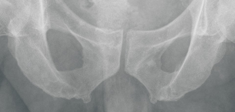
When the remains at St Thomaskirche were transferred to a new coffin in 1949, surgeon Wolfgang Rosenthal had the opportunity to inspect the skeleton. Although he did not notice any special features of the skull, he detected multiple bony outgrowths, which he considered to be exostoses, on the pelvic ring.3 He claimed His had mistaken these for signs of “arthrosis deformans” (osteoarthritis). He also noticed overdeveloped Muskellinien (“muscle lines”; bony outgrowths at sites of muscle and ligament attachment, now termed enthesophytes) on the arms of the skeleton. These outgrowths were assumed to be a sign of physical strength in people who use their arms powerfully over a long period. Rosenthal stated that multiple exostoses on the arms and legs are generally found in horseback riders, soldiers and sportsmen, in response to repetitive mechanical trauma. At that time, such formations were termed Reiter- und Exerzierknochen (“horseback rider and military drill bones”). A modern interpretation of these lesions is shown in Box 2.
In 2006, we presented a detailed scientific plan for examining the remains to the board of St Thomaskirche. A major goal was to compare DNA from the skeleton with DNA from one of Bach’s sons, Carl Philipp Emanuel (1714–1788), whose remains are kept in St Michaeliskirche in Hamburg, Germany.10 Unfortunately, the board of St Thomaskirche rejected our proposal, through which modern science might have shed more light on this intriguing riddle.
A second part of our investigation was to reproduce the research reported by Rosenthal. We undertook a study to explore the presence of enthesophytes in modern-day organists. Details are shown in Box 3.
Based on the available evidence, we believe it is unlikely that the the skeleton in question is that of Bach. The site of Bach’s grave was based on a doubtful oral tradition, and the value of the coffin being made of oak seems overestimated. Wilhelm His hoped his examination would find indicators of an extraordinarily developed musical talent, although a direct relationship between a highly developed peripheral sense of hearing and musical virtuosity has not been proven. A famous counterexample is Ludwig van Beethoven, who had hearing problems for half his life and wrote many compositions while completely deaf.12 Furthermore, the method His used to reconstruct Bach’s face was based on the assumption of a relationship between the skull and overlying soft tissue, which is disputed.13-15
2 Exostoses, enthesophytes and Reiterknochen
The latter are induced by long-term mechanical strain, both from forces external to the body, as in carrying heavy burdens, and from internal forces, such as hyperextension of the limbs.8 Examples include:
Reiterknochen and Exerzierknochen (myositis ossificans) — ossified muscle tissue caused by repetitive mechanical trauma. For example, soldiers who frequently present a rifle during drill may develop local ossification of muscle tissue in the left pectoralis. According to the modern definition, these are true extraskeletal bone formations in muscle tissue, and hence are seldom attached to bone.9
- Richard H C Zegers1,2
- Mario Maas1
- A (Ton) G Koopman3
- George J R Maat4
- 1 Academic Medical Center, Amsterdam, The Netherlands.
- 2 University of Amsterdam, Amsterdam, The Netherlands.
- 3 Faculty of Creative and Performing Arts, Leiden University, Leiden, The Netherlands.
- 4 Barge’s Anthropologica, Department of Anatomy, Leiden University Medical Center, Leiden, The Netherlands.
We thank the 12 church organists for their voluntary and enthusiastic cooperation, the hospitals that took their radiographs, and P J M van der Eerden, MD (Department of Radiology, Academic Hospital of Groningen, Groningen, The Netherlands) for commenting on the radiographs.
None identified.
- 1. His W. Anatomische Forschungen über Johann Sebastian Bach’s Gebeine und Antlitz nebst bemerkungen über dessen Bilder. Des XXII Bandes der Abhandlungen der mathematisch-physischen Classer der Königl. Sächsischen Gesellschaft der Wissenschaften. Leipzig: Hirzel, 1895.
- 2. His W. Johann Sebastian Bach. Forschungen über dessen Grabstätte, Gebeine und Antlitz. Bericht an den Rath der Stadt Leipzig. Leipzig: Vogel, 1895.
- 3. Rosenthal W. Die Identifizierung der Gebeine Johann Sebastian Bachs. Mit Bermerkungen über die “Organistenkrankheit”. Mitteilungen der Deutschen Akademie der Naturforscher. Leopoldina 1962/1963; 8/9: 235-241.
- 4. Wustmann G. Bach’s Grab. Grenzbote 1894; 42: 117-126.
- 5. Wustmann G. Die Auffindung der Gebeine Johann Sebastian Bachs. Grenzbote 1895; 54: 415-425.
- 6. Ulrich H. Johann Sebastian Bach. Ein historisches Rekonstruktionsverfahren auf dem Schädel. In: Schädel-Schicksale historischer Persönlichkeiten. Komponisten und Maler. München: Dr Friedrich Pfeil, 2004: 19-26.
- 7. Leopold D, Hammer HJ. Which picture characterizes JS Bach best? Craniofacial superimposition as a method of comparing the feature of the skull. Fingerprint Whorld 1989; 15: 61-63.
- 8. Kennedy KA. Skeletal markers of occupational stress. In: Iscan MY, Kennedy KA, editors. Reconstruction of life from the skeleton. New York: Alan R Liss Inc, 1989: 129-160.
- 9. Rogers J, Waldron T. A field guide to joint disease in archaeology. Chichester, UK: Wiley, 1994: 20-25.
- 10. Dreiling SA. Pompöser Leichenzug zur schlichten Grabstätte. Die vergessenen Toten im Gruftgewölbe der Hamburger St Michaelis-Kirche 1762-1813. Hamburg: Medien-Verlag, 2006.
- 11. Benjamin M, Toumi H, Ralphs JR, et al. Where tendons and ligaments meet bone: attachment sites (“entheses”) in relation to exercise and/or mechanical load. J Anat 2006; 208: 471-490.
- 12. Kubba AK, Young M. Ludwig van Beethoven: a medical biography. Lancet 1996; 347: 167-170.
- 13. Nelson LA, Michael SD. The application of volume deformation to three-dimensional facial reconstruction: a comparison with previous techniques. Forensic Sci Int 1998; 94: 167-181.
- 14. Maat GJ. Facial reconstruction. A review and comment. Talanta 1998-1999; 30-31: 247-253.
- 15. Stephan CN, Henneberg M. Recognition by forensic facial approximation: case specific examples and empirical tests. Forensic Sci Int 2006; 156: 182-191.
Abstract
A skeleton alleged to be that of Johann Sebastian Bach (1685–1750) was exhumed from a graveyard in Leipzig, Germany, in 1894, but its authenticity is not established.
In 1895, anatomist Wilhelm His concluded from his examination of the skeleton and reconstruction of the face that it most likely belonged to Bach.
In 1949, surgeon Wolfgang Rosenthal noticed exostoses on the skeleton and on x-rays of 11 living organists and proposed a condition, Organistenkrankheit, which he interpreted as evidence that the skeleton was Bach’s.
However, our critical assessment of the remains analysis raises doubts: the localisation of the grave was dubious, and the methods used by His to reconstruct the face are controversial.
Also, our study of the pelvic x-rays of 12 living professional organists failed to find evidence for the existence of Organistenkrankheit.
We believe it is unlikely that the skeleton is that of Bach; techniques such as DNA analysis might help resolve the question but, to date, church authorities have not approved their use on the skeleton.