




Volume 187 - Issue 8
Raw salmon or red herring: ascending paralysis with suspected seafood poisoning
Authors: Naren Gunja, Robert P Dowsett and Karl Ng
Med J Aust 2007; 187 (8): 468-469. || doi: 10.5694/j.1326-5377.2007.tb01359.x
Published online: 15 October 2007
Published online: 15 October 2007
A 16-year-old boy presented with rapidly progressive ascending paralysis 1 hour after eating raw salmon. Seafood poisoning was initially considered. Although salmon is not a common cause of toxic seafood poisoning, cases have been reported in the Pacific region. The patient rapidly developed acute left heart and respiratory failure, and investigations revealed a rare tracking intramedullary haematoma of the spinal cord. Structural abnormalities of the central nervous system may present with acute paralysis and spinal shock, mimicking toxicological syndromes.
Clinical record
A 16-year-old boy presented to the emergency department with severe epigastric pain, headache and ascending lower limb weakness 1 hour after eating raw salmon. The weakness ascended rapidly over the next hour to involve the upper limbs. In hospital, he was alert, but was soft-voiced and in obvious respiratory distress. He had tachycardia (heart rate, 110 beats per min), hypertension (blood pressure, 205/128 mmHg), tachypnoea (22 breaths per min) and oxygen saturation of 89% (using a non-rebreathing oxygen mask). Although his sensation and mental state appeared normal, he had profound quadriparesis. Pupils were 3 mm in diameter, equal and sluggishly reactive to light. Because of a rapid deterioration in his respiratory status, the patient was intubated, ventilated, and sedated with a propofol infusion before a more complete neurological examination could be completed.
The patient’s medical history included thalassaemia minor and a short viral illness 2 weeks previously. He denied using any regular medications or substance misuse.
Initial chest radiography revealed clear lung fields, but, 10 minutes after intubation, pinkish frothy sputum was noted from the endotracheal tube, and there were widespread crackles. An urgent echocardiogram showed severe global hypokinesis with a left ventricular ejection fraction estimated as less than 30%. Laboratory tests revealed neutrophilia, but all other results of a full blood count, serum electrolyte concentrations, renal function, liver function tests and coagulation screen were normal. Serum creatine kinase and creatine kinase-MB concentrations were raised, and peaked the following day at 2716 U/L (reference range [RR], 30–135 U/L) and 10 μg/L (RR, < 0.7 μg/L), respectively, suggesting myocardial injury. An electrocardiogram showed sinus rhythm with voltage criteria for left ventricular hypertrophy, but no acute ischaemic changes.
Provisional diagnosis at this stage was a neurological or toxicological aetiology. The fact that the patient’s mother also ate the salmon without becoming unwell counted against seafood poisoning but did not exclude it (eg, in puffer-fish poisoning, a specific part of the fish is most poisonous).
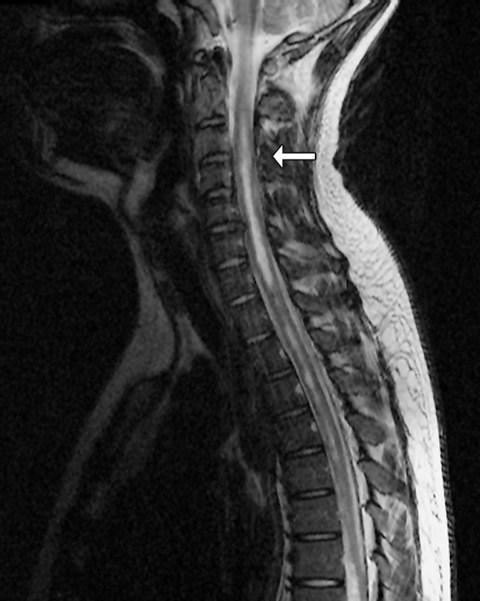
To exclude a cervical lesion, magnetic resonance imaging (MRI) of the spine and brain was performed urgently, with the intention of proceeding to nerve conduction studies if MRI results were normal. The MRI showed an extensive intramedullary haemorrhage within the spinal cord, originating at the T7/T8 vertebral level, and extending from the conus caudally to the cervicomedullary junction rostrally (Box 1).
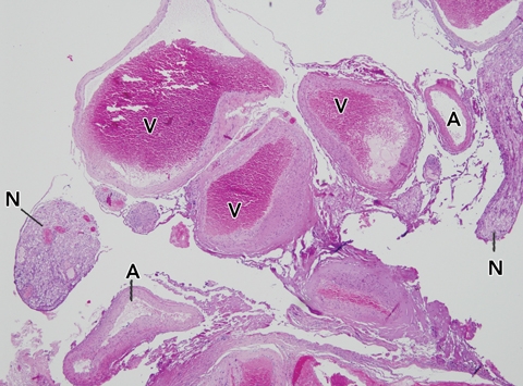
The patient was managed supportively in the intensive care unit, with complete resolution of pulmonary oedema and normalisation of cardiac function by Day 3. A spinal angiogram confirmed the presence of an arteriovenous malformation arising from the anterior spinal cord at the T9 vertebral level. The malformation was excised the following week, and histopathological examination of the resection specimen confirmed the radiological diagnosis (Box 2).
After 6 months of intensive rehabilitation, the patient had restored power in one arm, but remained paralysed below the T9 cord level.
Discussion
Initially, this case represented a diagnostic challenge, with acute onset, rapidly progressive ascending paralysis associated with left ventricular failure, and a history of seafood ingestion. MRI gave the correct diagnosis, obviating the need for peripheral neurophysiological investigations. Useful clues to the diagnosis of myelopathy, such as sphincter dysfunction and the level of sensory loss, were difficult to assess because of the patient’s sudden cardiorespiratory deterioration. The initial differential diagnosis is summarised in Box 3.
Toxicological considerations
Poisoning from ingested seafood is a global and increasing problem that should be considered in the emergency department. Seafood associated with medically important poisoning include puffer fish (fugu), ciguateric fish, several types of shellfish and mussels, as well as fish from the Scombridae family (eg, mackerel and tuna). Salmon is not known as a major cause of seafood toxin poisoning, although scombroid and ciguatera have been described after salmon ingestion.1-3
Tetrodotoxin from fugu fish causes a rapid descending paralysis and cardiovascular collapse in severe cases.4 Paralysis is uncommon in ciguatera, which is caused by a toxin produced by marine dinoflagellates.5 Paralytic shellfish poisoning is clinically similar to tetrodotoxin poisoning and typically causes a descending paralysis. Autonomic disturbance can also be a feature of marine neurotoxin poisoning.
Other toxins that can cause paralysis include botulinum, diphtheria and tick paralysis toxin, but these have slower onset of action and produce prodromal symptoms. Organophosphate poisoning is also worth considering: paralysis and copious bronchial secretions consistent with cholinergic toxicity are seen.
Neurological considerations
The hyperacute onset of our patient’s symptoms suggested an acute inflammatory demyelinating polyneuropathy, especially in view of the preceding infective illness. Even so, the presentation and progression of the weakness were unusually rapid. Similarly, autoimmune neuromuscular junction disorders, such as myasthenia gravis and Lambert–Eaton myasthenic syndrome, run a more subacute, relapsing course. Periodic paralysis can cause sudden weakness, but there is often a history of recurrent attacks. Toxic and inflammatory myopathies and neuropathic heavy metal poisoning have a more chronic and progressive course.
The cause in our patient was an intradural intramedullary arteriovenous malformation, the most common form of spinal cord vascular lesion in childhood and adolescence.6 These lesions can haemorrhage and cause catastrophic autonomic dysregulation accompanied by spinal shock. Complications include neurogenic pulmonary oedema, with the sympathetic storm contributing to transient myocardial impairment via direct neurogenic and humoral mechanisms.
Conclusion
This patient illustrates an uncommon scenario of rapidly progressive flaccid paralysis presenting to the emergency department. Emergency physicians need to consider a broad neurological and toxicological differential diagnosis in the patient with flaccid paralysis. Structural abnormalities of the central nervous system may present with acute paralysis and spinal shock, mimicking toxicological syndromes.
1 Magnetic resonance image of the cervical spinal cord
 |
|
A sagittal T2-weighted image showed a high signal (arrow) within the cervical cord caused by blood breakdown products (intracellular methaemoglobin). |
2 Lesion excised from the spinal cord
3 Toxicological and neurological causes of acute paralysis
Toxicological: tick paralysis (Ixodes spp.)
Neurological (peripheral): acute inflammatory demyelinating polyradiculoneuropathy (Guillain–Barré syndrome)
Toxicological: puffer-fish (tetrodotoxin) poisoning, paralytic shellfish poisoning, snake bite, botulism, diphtheria
Neurological (peripheral): myasthenia gravis
Toxicological: ciguatera, neurotoxic shellfish poisoning, organophosphate poisoning
Neurological (peripheral): myopathies (including myositis and periodic paralysis), acute poliomyelitis and toxic neuropathies
Competing interests
None identified.
Acknowledgements
We thank Dr D Brazier and Dr J Brewer, Royal North Shore Hospital, for the magnetic resonance image and histopathological image, respectively.
References
- Dowsett RP. Seafood toxins. In: Wolfson AB, Hendey GW, Hendry PL, et al, editors. Harwood-Nuss’ clinical practice of emergency medicine. 4th ed. Philadelphia: Lippincott Williams and Wilkins; 2005: 1710-1714. 0_BIHCGHHC
- Smart DR. Scombroid poisoning: a report of seven cases involving the Western Australian salmon, Arripis truttaceus. Med J Aust 1992; 157: 748-751. 0_pgfId-1094740
- Ebesu J, Nagai H, Hokama Y. The first reported case of human ciguatera possibly due to a farm-cultured salmon. Toxicon 1994; 32: 1282-1286. 0_i1091872
- Isbister GK, Kiernan MC. Neurotoxic marine poisoning. Lancet Neurol 2005; 4: 219-228. 0_i1091874
- Swift AE, Swift TR. Ciguatera. J Toxicol Clin Toxicol 1993; 31: 1-29. 0_i1091876
- Krings T, Mull M, Gilsbach JM, Thron A. Spinal vascular malformations. Eur Radiol 2005; 15: 267-278. 0_i1091878
Motor Neuron Disease Mortality Trends in Australia From 1986 to 2023: A Population-Based Study
Carol M. Y. Lee, Rupendra N. Shrestha, Julian Gold, Mark Stevenson, Kelly L. Williams, Lyndal Henden, Sandrine K. K. Chan Moi Fat, Zoe Zussa, Amanda Wright, Martin Jude, Mark A. S. Laidlaw, Willem J. R. Fokkink, Dominic B. Rowe
Mild traumatic brain injury and concussion and persisting post‐concussion symptoms: new guidelines to support evidence‐based assessment and management in Australia and Aotearoa New Zealand
Karen M Barlow, Jennie L Ponsford, Alice Theodom, Gill Cowen, Gavin A Davis, Vicki Anderson, Franz E Babl, David Cole, Jennifer Cullen, Stuart R Dalziel, Melinda Fitzgerald, Howard Flavell, Caroline Yates, Rebecca Kimble, John H Olver, Rhonda Orr, Mark Ralfe, Michael Rose, Nick Rushworth, Julia Treleaven, Gary Browne, Nathan Delang, Sarah Harris, Gary Mitchell, Sean Tweedy
The prevalence of and variation in indicators of the quality and safety of long term aged care in Australia, 2019: a cross‐sectional population‐based study
Tesfahun C Eshetie, Gillian E Caughey, Catherine Lang, Olivia Ryan, Renuka Visvanathan, Craig Whitehead, Keith Evans, Janet K Sluggett, Jyoti Khadka, Carolyn Dawkins, Helena Williams, Miranda Starke, Sara Blunt, Anne Liddell, Megan Corlis, Anna Sheppeard, Penelope Lello, Marilyn Thien, Steven L Wesselingh, Maria C Inacio
Health and economic benefits of improving pre‐hospital identification of stroke in Australian women: a modelling study
Thomas Gadsden, Lei Si, Emily R Atkins, Cheryl Carcel, Xia Wang, Stephen Jan, Mark Woodward, Laura E Downey
Symptom burden, quality of life, and diagnostic journey of people with postural orthostatic tachycardia syndrome, Australia, 2021–24: a descriptive patient registry data study
Marie‐Claire Seeley, Gemma Wilson, Eric Ong, Amy Langdon, Jonathan Chieng, Danielle Bailey, Kristina Comacchio, Amanda Page, Dennis H Lau, Celine Gallagher
The intersection of rurality and dementia prevalence in Australia for Aboriginal and Torres Strait Islander and non‐Indigenous peoples
Antonia J Clarke, Marwan Ahmed, Judith M Katzenellenbogen, John Towney (Wiradjuri), Anna H Balabanski, Adrienne Withall (Dharawal Yuin), Kylie Radford, Amy Brodtmann