




Volume 186 - Issue 6
Pulmonary artery aneurysm
Authors: Zubair Ahmad, Imrana Masood and Saurabh K Singh
Med J Aust 2007; 186 (6): 314. || doi: 10.5694/j.1326-5377.2007.tb00908.x
Published online: 19 March 2007
Published online: 19 March 2007
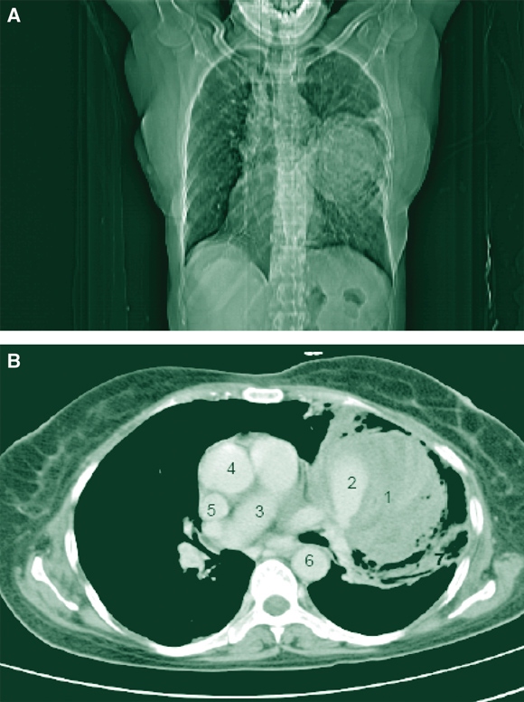
A 55-year-old woman was admitted to hospital with a 1-year history of recurrent haemoptysis. A chest radiograph showed a well defined, rounded left hilar mass with surrounding air-space shadowing (Box, A). Contrast-enhanced computed tomography revealed a thrombosed aneurysm of the left main pulmonary artery that extended into its branches, with surrounding lung consolidation (Box, B). Echocardiography excluded any valvular lesions or shunt. Tests for tuberculosis and HIV were negative, and antinuclear antibody levels were not raised. The aneurysm was thought to be idiopathic. It was surgically resected, and the postoperative course was uneventful.
Fatty Liver Disease in Australia: A Narrative Review on the Epidemiology, Natural History, Prognostication and Management in People With Metabolic Dysfunction
Karl Vaz, Daniel Clayton-Chubb, William W. Kemp, Stuart K. Roberts, Ammar Majeed
Sex Differences in Management and Outcomes of People With ST-Elevation Myocardial Infarction, New South Wales, 2011–2020: A Retrospective Cohort Study
Adam C. Livori, Jedidiah I. Morton, Dianna J. Magliano, Jonathan E. Shaw
Rethinking Rheumatic Fever Diagnosis: Progress, Pitfalls and the Path Towards a Definitive Test
Anna P. Ralph, Rachel Webb, Tom Parks, Nicole J. Moreland, Asha C. Bowen, Benjamin Jones, Emma Ndagire, Andrea Z. Beaton
Sex Differences in Management and Outcomes of People With ST-Elevation Myocardial Infarction, New South Wales, 2011–2020: A Retrospective Cohort Study
Samia Kazi, Simone Marschner, Clara K. Chow
Distribution of Cardiovascular Disease Risk Based on the Updated 2023 Guideline-Recommended Australian Cardiovascular Disease Risk Algorithm and Comparison With the 2012 Algorithm: An Observational Study
Nina Lazarevic, Meghana Bhat, Grace Joshy, Danielle C. Butler, Mark Woodward, Anushka Patel, Rod T. Jackson, Garry Jennings, Rosemary Wyber, Ellie Paige, Emily Banks
They Brought the Hospital to Me
Hugh N. Burrill