




Volume 185 - Issue 7
Eosinophilic oesophagitis
Authors: Alyson Kakakios and Ralf G Heine
Med J Aust 2006; 185 (7): 401. || doi: 10.5694/j.1326-5377.2006.tb00620.x
Published online: 2 October 2006
Published online: 2 October 2006
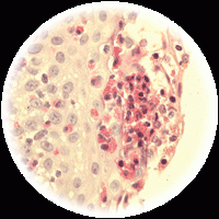
Eosinophilic oesophagitis (EO), an isolated eosinophilic inflammation of the oesophagus, is the most common of the eosinophilic gastrointestinal disorders. EO is of increasing clinical significance in many developed countries,1 in parallel with the recent increase in food allergic disorders. A retrospective study of Western Australian children reported a dramatic rise in prevalence of EO (from 0.05 cases per 10 000 children in 1995 to 0.89 cases per 10 000 children in 2004) (Level III-2).2 The estimated prevalence in the US population aged 0–19 years is even higher, at 4.3 cases per 10 000 individuals.3
Clinical features. Patients present with symptoms indistinguishable from those of gastro-oesophageal reflux (GOR); however, unlike people with GOR, they are generally unresponsive to treatment with proton pump inhibitors (Level III-2). Infants often have additional clinical features, including feeding difficulties, feeding refusal and/or poor weight gain. In older children and adults, oesophageal food impaction is the most characteristic symptom (Level III-3), and should alert clinicians to include EO in the differential diagnosis. Typically, patients with EO have associated atopic disorders, including asthma and eczema.
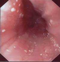
Cause. The cause of EO is not clear, but the condition is closely associated with atopic disorders, and there is evidence that both IgE- and non-IgE-mediated food allergy are involved in its aetiology. Foods commonly implicated include cows milk, soy, wheat and egg.3 Data from animal experiments and anecdotal clinical observations suggest that inhalant allergens may also contribute.
In keeping with murine models, recent gene array studies in people with EO have shown increased gene expression for eotaxin-3, a chemokine that promotes the migration of eosinophils into the oesophagus.1
Diagnosis. The diagnostic hallmark of EO is a dense, eosinophilic infiltrate involving the entire oesophageal mucosa, which is normally free of eosinophils. Key diagnostic criteria are basal layer hyperplasia and the presence of more than 20 eosinophils per high power field (× 400 magnification) in gastroscopic biopsies of the lower and upper oesophagus (Level III-3). Although oesophageal eosinophils are also seen in patients with reflux oesophagitis, mucosal eosinophil counts in such patients are lower (< 5 per high power field), and the eosinophils are limited to the lower oesophagus. There is a typical mucosal appearance in many patients with EO (thickened mucosa, with longitudinal furrowing and superficial white plaques [Box]) — although in about a third of patients the mucosa will look macroscopically normal.
Skin prick testing (SPT) and atopy patch testing (APT) (the application of food or food extracts to the skin for 48 hours) are thought to be helpful in identifying potential causative food allergens,3 but prospective studies are needed to evaluate their predictive value.
Management. To date, no completed randomised controlled trials evaluating the benefits and adverse effects of medical treatments for EO are available.
Infants and young children may respond to dietary allergen restriction or an elemental diet. A diagnostic trial of an amino acid-based formula (AAF) for 6–8 weeks may be useful in determining whether a patient is diet-responsive. The treatment response needs to be assessed by repeat endoscopy, as not all patients will improve. If remission is achieved, food items can be gradually reintroduced into the diet, as tolerated, taking SPT and APT findings into consideration.3
In older children and adults, an elemental diet is often impractical and poorly tolerated due to the taste of AAF. These patients can be treated with swallowed topical corticosteroid aerosols (fluticasone) or, in refractory cases, with systemic corticosteroids.1 Other drugs, including cromoglycate and montelukast, have been used in uncontrolled trials, but their efficacy is poorly documented. Novel monoclonal antibodies against interleukin-5 may also be of benefit in treating EO, but prospective studies are not yet available.
Evidence-based practice tip
Infants and young children with eosinophilic oesophagitis often respond to an elemental diet, while older children and adults have been successfully treated with swallowed topical corticosteroid aerosols (fluticasone) (Level III-2).*
Prognosis. The long-term prognosis for EO is largely unknown. In some infants and young children with food protein-induced EO, the disease may remit due to development of oral tolerance to the offending food protein. However, EO usually follows a chronic relapsing course. To date, no studies have shown an increased risk of malignancy in patients with EO, but there is evidence that uncontrolled chronic eosinophilic inflammation may cause subepithelial fibrosis and remodelling, which eventually may cause obstructive dysphagia, strictures or persistent oesophageal narrowing.
References
- Furuta GT, Straumann A. Review article: the pathogenesis and management of eosinophilic oesophagitis. Aliment Pharmacol Ther 2006; 24: 173-182.
- Cherian S, Smith NM, Forbes DA. Rapidly increasing prevalence of eosinophilic oesophagitis in Western Australia. Arch Dis Child 2006 Jul 28; [Epub ahead of print]. 0_i1091815
- Spergel JM, Andrews T, Brown-Whitehorn RF, et al. Treatment of eosinophilic esophagitis with specific food elimination diet directed by a combination of skin prick and patch tests. Ann Allergy Asthma Immunol 2005; 95: 336-343. 0_i1091817
Rhabdomyolysis After COVID-19
Navya Kataria, Abel Devadass, Lana Sundac, Laura De Souza, Nabeel Sheikh, Samantha Ng, Scott Campbell, Yishen Wang, Ross S. Francis
Technological advances in the search for a CURE for asthma
Sara Quon, Timothy M Johanson, Ridhima Wadhwa, Alen Faiz, Anthony Flynn, Gary P Anderson, Amanda J Cox, Nicholas P West, Michael P Menden, Rhys S Allan
The CURE Asthma roadmap
Gary P Anderson, Anthony Flynn, Phil G Bardin, John D Blakey, Shyamali C Dharmage, Paul Foster, Peter G Gibson, Adam Jaffe, Alan James, Christine R Jenkins, Sundram Sivamalai, Peter D Sly, Guy B Marks, Vanessa M McDonald, Judy Wetttenhall
The first live term birth following uterus transplantation in Australia
Rebecca Deans, Brigitte Gerstl, Antonia W Shand, Sarah Lyons, Aaron Budden, Helen L Barrett, Grant Luxton, Mangalee Fernando, Kenneth Yong, Karen Keung, Kaushalya Arulpragasam, Henry Pleass, King Man Wan, Eva Kehag, Jana‐Emily Pittman, Mianna Lotz, Maria Fenn, Erin Nesbitt‐Hawes, Lily Byun, Katrina Tang, Mats Brannstrom, Jason Abbott
The public health impacts of mining in Australia
Javier Cortes‐Ramirez, Ruby N Michael, Leisa‐Maree Toms, Melissa Haswell
Lingual Raynaud phenomenon
Michael Taggart, Mina John