




Volume 182 - Issue 12
Potential pitfalls in the diagnosis of phaeochromocytoma
Authors: Jane L Harding, Michael W Yeh, Leigh W Delbridge, Stan B Sidhu and Bruce G Robinson
Med J Aust 2005; 182 (12): 637-639. || doi: 10.5694/j.1326-5377.2005.tb06852.x
Published online: 20 June 2005
Published online: 20 June 2005
Six patients being evaluated for phaeochromocytoma had misleading investigative findings: all initially had raised urinary catecholamine levels, and five had adrenal masses on imaging studies. Adrenalectomy in these five patients revealed only one pathologically confirmed phaeochromocytoma. Tricyclic antidepressant use produced misleading elevations in urinary catecholamine levels in three patients. 24-hour urine studies should be performed at least twice, after eliminating confounding factors (stressors, medications).
Clinical record
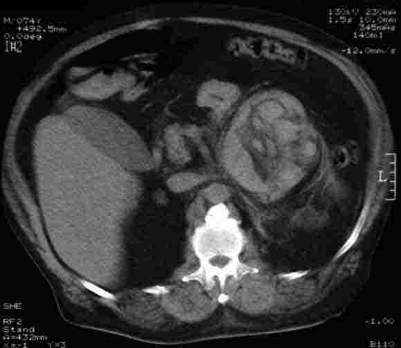
The details of six patients treated in the period April 1999 – October 2003 by members of the Section of Endocrine Surgery of the Royal Australasian College of Surgeons are outlined in Box 1. In all patients, clinical suspicion of phaeochromocytoma was raised by the presence of hypertension, paroxysmal symptoms, or both. Twenty-four-hour urinary catecholamine levels were found to be elevated, although in Patients 1–5 these abnormalities were confined to one or two analytes only. In all except Patient 2, adrenal lesions were discovered on imaging, with large haemorrhagic masses detected in patients with an acute presentation (Patients 5 and 6). Patients with abnormalities on computed tomography (CT) underwent surgery. In Patients 1, 3, and 4, the excised tissues were found to be pathologically normal or to show mild enlargement (benign). In Patients 5 and 6, blood clot and necrotic tissue comprised the bulk of the specimens (Box 2), with a phaeochromocytoma discovered in the latter patient. Of note, Patients 1–3 were receiving tricyclic antidepressants for non-standard uses that did not include the treatment of major depression.
Discussion
False positive biochemical test results for phaeochromocytoma are common and present particular problems because of the low prevalence of the disease. The reported incidence of phaeochromocytoma is 2–8 per million people annually, accounting for less than 0.2% of all patients with hypertension.1 Despite the fact that phaeochromocytoma is a rare cause of hypertension, the diagnosis merits consideration in a potentially large group of patients for two reasons. Firstly, although the disease is frequently fatal if unrecognised, surgical removal is highly effective, achieving cure in greater than 90% of cases.2 Secondly, because no array of clinical indicators has proven to reliably include or exclude phaeochromocytoma,3 physicians must maintain a high level of suspicion and consider biochemical testing in patients at risk. The pretest probability for phaeochromocytoma is close to 0.5% (1 in 200 patients tested) in the presence of hypertension and suggestive symptoms.4 Assuming a specificity of 85% for biochemical testing, 30 false positive results are generated for every one patient with phaeochromocytoma identified.5
Fortunately, most false positive tests can be unmasked with careful additional investigation and/or the elimination of factors known to confound biochemical tests for levels of catecholamines and their metabolites.
The misleading elevations in urinary noradrenaline levels in Patients 1–3 can be ascribed to their taking tricyclic antidepressants. Medications and conditions that may result in raised levels of plasma and/or urine catecholamines and their metabolites, and result in false positive test results for phaeochromocytoma, are listed in Box 3. Among these drugs, tricyclic antidepressants and phenoxybenzamine have been the most frequently implicated, together accounting for more than 40% of medication-associated false positive results in a recent large study.6
In Patients 3 and 4, false positive biochemical findings led to the identification of small (1.5 cm) adrenal masses on CT scanning, both of which were found, on histopathological examination, to be benign. Clinically unapparent adrenal masses (“incidentalomas”) are found in 2.1% of subjects at autopsy and in 1%–4% of abdominal imaging studies. Most of these masses are benign, hormonally inactive tumours that do not require surgical management.7
Phaeochromocytomas presenting with acute haemorrhage at presentation have been reported previously,8,9 with haemorrhagic tumours often losing characteristic imaging appearances and functional markers. Tumour necrosis may initially result in massive catecholamine release, followed by failure to demonstrate excess catecholamine levels, as was seen in Patient 6. Mildly elevated urinary catecholamine levels may also be seen as a consequence of hyperadrenergia from an acute stress response at the time of haemorrhage into a non-phaeochromocytoma lesion, as occurred in Patient 5.
Role of biochemical testing
Although measurement of plasma free metanephrine levels has been recently advocated by some groups, 24-hour urinary catecholamine levels and total metanephrine level have consistently proven to be the most specific tests available for the diagnosis of phaeochromocytoma.5,10 Elevations in the level of one or more of these analytes (above the 95% reference range designated as the upper limit of normal by laboratories) are common in patients with paroxysmal symptoms or poorly controlled hypertension not due to phaeochromocytoma. Thus, we recommend that higher cut-off values, roughly two times the upper limit of normal for most laboratories, be used to identify patients suitable for further workup.
Repeat biochemical testing 6 weeks after stopping drugs likely to confound the results is ideal, and tests performed during major physical or psychological stress should be interpreted with extreme caution (if performed at all). It is important to note that alterations in plasma catecholamine levels may be caused not only by medications, but also by the underlying diseases being treated (eg, major depression in the case of tricyclic antidepressants or severe heart disease in the case of β-blockers).11-13
All patients should undergo at least two 24-hour urine collections for measuring levels of catecholamines and their metabolites. Clonidine suppression testing — the measurement of plasma free normetanephrine before and after the oral administration of 0.3 mg clonidine — is highly sensitive and specific, and may be a useful adjunct in patients with more than one prior set of equivocal tests.6
Role of imaging
Whether imaging studies (both anatomical and functional) play a role in diagnosing phaeochromocytoma, as opposed to only localising tumours already diagnosed biochemically, remains controversial. In the patients described above, 131I‑metaiodobenzylguanidine scanning yielded true negative results in Patients 1–3, consistent with its known high specificity.4 However, current evidence suggests that, when appropriate biochemical tests are used, little discriminatory value is to be gained from imaging,14 and our experience illustrates how incidental radiographic findings may lead to unnecessary surgery.
1 Summary of the clinical records of six patients investigated for phaeochromocytoma
Patient age/sex |
Presentation |
Blood pressure (mmHg) |
Medications |
24-h Urinary catecholamine levels (nmol/d or μmol/d)* |
Radiological investigation† |
Surgical findings/ Clinical course |
Pathology |
||||||||
Patient 1 49/M |
Weight loss (5 kg in 3 months), paroxysmal anxiety attacks, drenching sweats, chronic right flank pain |
124/80 |
Clomipramine, 50 mg/day (anxiety) |
Adrenaline, 91/84 Noradrenaline, 860/1224 |
CT: 3 x 6-cm mass abutting upper pole of right kidney MIBG: negative |
Lobulated upper pole of kidney due to right renal artery branch running within posterior cleft |
Normal adrenal tissue |
||||||||
Patient 2 41/M |
Migraines, chronic back pain, worsening hypertension, palpitations |
145/85 |
Amitriptyline, 150 mg/day (migraine prophylaxis); amlodipine, 5 mg/day; ramipril, 5 mg/day; indapamide, 2.5 mg/day |
Adrenaline, 7/12 Noradrenaline, 485/1295 |
CT: No abnormalities MIBG: No abnormalities |
Normal findings on repeat urine studies after stopping amitriptyline |
— |
||||||||
Patient 3 64/M |
Metastatic prostate cancer, poorly controlled hypertension |
180/95 |
Imipramine, 100 mg/day (neuropathic pain); nifedipine, 180 mg/day; ramipril, 5 mg/day; chlorothiazide, 1000 mg/day |
Adrenaline, 115/99 Noradrenaline, 1070/1130 VMA, 33/38 |
CT: 1.5-cm left adrenal mass MIBG: negative |
Small left adrenal mass, macroscopically consistent with an adenoma |
Benign adrenal adenoma |
||||||||
Patient 4 76/F |
Chronic hypertension, 15-month history of paroxysmal nausea and vomiting |
144/80 |
Captopril, 25 mg/day |
Adrenaline, 65/129 Noradrenaline, 371/451 Dopamine, 1.55/2.02 |
CT: 1.5-cm left adrenal mass |
Smooth lesion palpable within left adrenal gland |
Enlarged adrenal gland (10.2 g; reference, 4 g) with thickened cortex but normal medulla |
||||||||
Patient 5 75/M |
Sudden onset of intense back and left loin pain, chronic hypertension, weight loss (22 kg in 6 months), atrial fibrillation, diabetes, polymyalgia rheumatica |
150/90 |
Warfarin, 5 mg/day; captopril, 25 mg/day; digoxin, 0.25 mg/day; isosorbide mononitrate, 60 mg/day; verapamil, 240 mg/day; prednisone, 5 mg/day; metformin, 2 g/day; rosiglitazone, 4 mg/day; thyroxine, 0.125 mg/day |
Adrenaline, 250 Noradrenaline, 720 Metanephrine, 1.68 Normetanephrine, 2.49 |
CT: 12-cm heterogeneous left adrenal mass (Box 2) |
Large blood clot with associated desmoplastic reaction occupying most of left adrenal gland |
Myelolipoma with haemorrhagic fat necrosis |
||||||||
Patient 6 58/M |
Acute abdominal pain and hypertension, otherwise healthy |
210/95 |
— |
Adrenaline, 2426/51 Noradrenaline, 18 370/464 Metanephrine, 13.3/0.8 Normetanephrine, 35.5/3.6 |
CT: 5-cm haemorrhagic left adrenal mass MRI (2 weeks after presentation): subacute haemorrhage into area without any distinct adrenal mass |
12-cm dumbbell-shaped mass |
3-cm phaeochromo-cytoma, large organising blood clot and necrotic tissue |
||||||||
* Values separated by a forward slash represent separate collections, with abnormal values in bold. Reference ranges: adrenaline, < 100 nmol/d; noradrenaline, < 680 nmol/d; metanephrine, < 2.1 μmol/d; normetanephrine, < 5.6 μmol/d; VMA (vanillylmandelic acid), < 40 nmol/d; dopamine, < 3.0 μmol/d. |
|||||||||||||||
3 Medications and conditions that may cause false positive results of biochemical tests for phaeochromocytoma
Medication or condition |
Test(s) confounded |
||||||||||||||
Tricyclic antidepressants |
Urinary catecholamines and metanephrines, plasma free metanephrines |
||||||||||||||
Clozapine |
Urinary catecholamines and metanephrines |
||||||||||||||
Phenoxybenzamine |
Plasma free metanephrines |
||||||||||||||
Calcium channel blockers |
Plasma noradrenaline, urinary noradrenaline, urinary adrenaline |
||||||||||||||
β-adrenergic blockers |
Urinary catecholamines and metanephrines, plasma free metanephrines (minor effect) |
||||||||||||||
α1-adrenergic blockers |
Urinary noradrenaline |
||||||||||||||
Sympathomimetics |
Urinary catecholamines and metanephrines, plasma free metanephrines |
||||||||||||||
Buspirone |
Urinary metanephrines |
||||||||||||||
Major physical or psychological stress* |
Urinary catecholamines and metanephrines, plasma free metanephrines |
||||||||||||||
* Hypoglycaemia, hypoxia, hypovolaemia, stroke, surgery, myocardial infarction, heart failure, severe pain, depression, panic disorder, sleep apnoea. |
|||||||||||||||
Competing interests
None identified.
Acknowledgements
We thank Dr Maxwell Coleman (St Vincent’s Hospital, Sydney) and Dr Lionel Hartley (Princess Alexandra Hospital Endocrine Unit, Sydney) for their contribution of cases.
References
- O’Connor DT. The adrenal medulla, catecholamines and pheochromocytoma. In: Goldman L, Bennett JC, editors. Cecil textbook of medicine. Philadelphia, PA: WB Saunders Company, 2000: 1259.
- Manger WM, Eisenhofer G. Pheochromocytoma: diagnosis and management update. Curr Hypertens Rep 2004; 6: 477-484. i1086028
- Stein PP, Black HR. A simplified diagnostic approach to pheochromocytoma. A review of the literature and report of one institution’s experience. Medicine (Baltimore) 1991; 70: 46-66. i1086030
- Pacak K, Linehan WM, Eisenhofer G, et al. Recent advances in genetics, diagnosis, localization, and treatment of pheochromocytoma. Ann Intern Med 2001; 134: 315-329. i1086032
- Kudva YC, Sawka AM, Young WF Jr. Clinical review 164: The laboratory diagnosis of adrenal pheochromocytoma: the Mayo Clinic experience. J Clin Endocrinol Metab 2003; 88: 4533-4539. i1086034
- Eisenhofer G, Goldstein DS, Walther MM, et al. Biochemical diagnosis of pheochromocytoma: how to distinguish true- from false-positive test results. J Clin Endocrinol Metab 2003; 88: 2656-2666. i1086036
- Grumbach MM, Biller BM, Braunstein GD, et al. Management of the clinically inapparent adrenal mass (“incidentaloma”). Ann Intern Med 2003; 138: 424-429. i1086038
- Jacobs LM, Williams LF, Hinrichs HR. Hemorrhage into a pheochromocytoma. JAMA 1978; 239: 1156. i1086040
- Lehman DJ Jr, Rosof J. Massive hemorrhage into an adrenal pheochromocytoma. N Engl J Med 1956; 254: 474-476. i1086042
- Lenders JW, Pacak K, Walther MM, et al. Biochemical diagnosis of pheochromocytoma: which test is best? JAMA 2002; 287: 1427-1434. i1086044
- Krentz AJ, Mikhail S, Cantrell P, Hill GM. Drug points: pseudophaeochromocytoma syndrome associated with clozapine. BMJ 2001; 322: 1213. i1086046
- Swedberg K, Bristow MR, Cohn JN, et al. Effects of sustained-release moxonidine, an imidazoline agonist, on plasma norepinephrine in patients with chronic heart failure. Circulation 2002; 105: 1797-1803.
- Veith RC, Lewis N, Linares OA, et al. Sympathetic nervous system activity in major depression. Basal and desipramine-induced alterations in plasma norepinephrine kinetics. Arch Gen Psychiatry 1994; 51: 411-422. i1086049
- Neumann HP. Imaging vs biochemical testing for pheochromocytoma. JAMA 2002; 288: 314-315 (author reply: 315). i1086051
Fatty Liver Disease in Australia: A Narrative Review on the Epidemiology, Natural History, Prognostication and Management in People With Metabolic Dysfunction
Karl Vaz, Daniel Clayton-Chubb, William W. Kemp, Stuart K. Roberts, Ammar Majeed
Sex Differences in Management and Outcomes of People With ST-Elevation Myocardial Infarction, New South Wales, 2011–2020: A Retrospective Cohort Study
Adam C. Livori, Jedidiah I. Morton, Dianna J. Magliano, Jonathan E. Shaw
Rethinking Rheumatic Fever Diagnosis: Progress, Pitfalls and the Path Towards a Definitive Test
Anna P. Ralph, Rachel Webb, Tom Parks, Nicole J. Moreland, Asha C. Bowen, Benjamin Jones, Emma Ndagire, Andrea Z. Beaton
Sex Differences in Management and Outcomes of People With ST-Elevation Myocardial Infarction, New South Wales, 2011–2020: A Retrospective Cohort Study
Samia Kazi, Simone Marschner, Clara K. Chow
Distribution of Cardiovascular Disease Risk Based on the Updated 2023 Guideline-Recommended Australian Cardiovascular Disease Risk Algorithm and Comparison With the 2012 Algorithm: An Observational Study
Nina Lazarevic, Meghana Bhat, Grace Joshy, Danielle C. Butler, Mark Woodward, Anushka Patel, Rod T. Jackson, Garry Jennings, Rosemary Wyber, Ellie Paige, Emily Banks
They Brought the Hospital to Me
Hugh N. Burrill