


Thorough assessment and adequate pain relief facilitates management of minor childhood injuries
Minor injuries in children are extremely common. The combination of a developing physical ability, lack of recognition of dangerous situations and a willingness to robustly explore their environment means that children are more likely than adults to injure themselves. Most of these injuries are managed by the child’s parents or carers, or by the child alone, and do not come to medical attention; it is not known how frequently these injuries occur.
Among children of all ages who attend an emergency department, a fall is the commonest cause of injuries. The next most common cause is a strike or collision with a blunt object.1 The most common types of injuries that children sustain are soft-tissue bruising, abrasions, lacerations, fractures and minor head injuries. The first three types of injuries will be dealt with here, and fractures and minor head injuries will be covered in the next article in the MJA Practice Essentials Series – Paediatrics (Fractures and minor head injuries: minor injuries in children II. Med J Aust 2005; 182 [20 June]).
Case study — a 13-month-old boy with a laceration to the head
A mother brings her normal, healthy, 13-month-old son to you 1 hour after he has fallen and struck the left side of his forehead on the edge of a glass coffee table, sustaining a laceration (Figure a). Management
|
|||||||||||||||
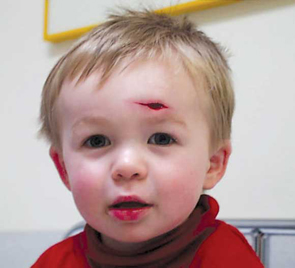 a: An open laceration to the forehead. |
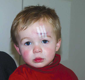 b: Wound closed by tissue adhesive and adhesive tape. |
||||||||||||||
The commonest sites of soft-tissue bruising in children are the head and lower legs. As bruising generally occurs at or near the site of trauma, the location can assist the clinical examination; for example, a large bruise to the occiput makes it necessary to exclude an intracranial injury; and, similarly, bruising of the fingers will raise suspicion of a fracture. The extent of the bruising, however, is not a particularly good indication of the amount of force sustained, as commonly there is impressive bruising and few, if any, other clinical signs.
It is important to consider the pattern of bruising and whether it is consistent with the history of trauma. Otherwise, it may raise the possibility of non-accidental injury. For example, multiple small rounded bruises occurring on the inner aspect of the upper arms are suggestive of the child being gripped by a hand in this area. Similarly, linear bruises across the buttocks may be caused by the child being struck with an object.
If bruising is extensive, has a typical pattern or occurs in the absence of trauma, or with only minor trauma, the child may have a condition such as idiopathic thrombocytopenic purpura, Henoch Schönlein purpura, or another bleeding disorder.
Management of bruising: A cool pack and oral analgesia are helpful in the first few hours after bruising; however, specific treatment is rarely necessary. Early mobilisation assists in a speedy recovery.
Lacerations are caused by a blunt force tearing tissues. They commonly occur as a result of a fall and are often situated on the scalp or over the bony prominences of the face. Incised wounds, caused by a sharp object such as glass or a knife cutting the skin, tend to occur on the hands or feet.
A number of therapeutic options are possible.2,3 Select the appropriate agent depending on the wound, the degree of pain, the experience of the staff, and the procedure that is likely to be performed. A summary of options are listed in Box 1.
Some agents such as morphine provide both analgesia and sedation. Be generous with the provision of analgesia and allow time for it to work, as poor pain control is one of the major causes of procedural failure in children.
Supplement pharmacological agents with distraction and guided imagery. Distract the child by focusing their attention on toys or objects, events such as sports or their favourite television program. Ask the child to imagine a pleasant place or event and encourage them to describe it to you.
Local anaesthesia options are also listed in Box 1.
Topical anaesthesia is painless, easy to apply and has a similar efficacy to infiltrated lignocaine.5 ALA (an adrenaline/lignocaine/amethocaine mixture) is a clear liquid that is applied topically by flooding directly into the wound. A cotton wool pledget inserted between the wound edges and soaked in ALA improves retention of the ALA fluid. It takes around 45 minutes to achieve maximal effect and has the added advantage of reducing bleeding. At present in Australia it is only available for use in hospitals.
EMLA (an eutectic mixture of lignocaine and prilocaine) is a cream that is usually applied to intact skin; however, it appears to be safe and effective for simple extremity lacerations even though it is not licensed for this use.6,7
Regional nerve blocks, such as digital, ulna or femoral nerve blocks, are very effective in children. A number of reference books are available that provide the anatomical knowledge and practical instruction necessary to perform them.3
All dirt and foreign material in the wound must be located and removed before closure. Superficial wounds may be safely cleaned with good quality tap water.8 Preparations such as aqueous chlorhexidine are painful to apply and of doubtful benefit. Irrigation with saline under pressure (using a 19-gauge needle on a 10–20 mL syringe) is a good way of dislodging and removing foreign material.
X-rays are helpful for detecting some foreign bodies, especially glass or metallic fragments. Wounds that require exploration should be anaesthetised first to allow more thorough examination and cleaning.
Small superficial wounds with opposed edges do not require closure and can be managed with dressings alone. Other wounds may be closed with tissue adhesives, adhesive strips, sutures or a combination of two or three of these.
Tissue adhesives are most successful on wounds that are less than 3 cm long, have clean straight edges, do not require deep sutures and are not under tension when the edges are opposed. They do not require local anaesthesia and are quick and easy to apply. Parents are usually extremely relieved to learn that this is an option for their child’s wound. The cosmetic result for a wound closed with tissue adhesives is the same as for wound closure achieved with sutures, staples or adhesive strips.9 However, with tissue adhesives (compared with sutures), there is a small increase in the incidence of wound dehiscence, but all other wound complications appear to be the same for both wound closure methods.9
Any area of the skin may be glued; however, gluing in the vicinity of the eye requires extreme care to prevent any glue dripping into the eye or on to the eyelashes. If gluing the scalp, remove any hair from the wound but do not shave or cut the surrounding hair. Before gluing, the wound must be dry and not bleeding — applying ALA first may assist with this.
To apply tissue adhesive, position the child so the wound is uppermost to minimise the glue running. Ensure the operator is wearing gloves; this is not only for cleanliness, but to ensure that it is the glove not the operator that is stuck to the child, if adhesive inadvertently runs on to the operator’s fingers. The hand can then be removed from the glove, and the glove fingers cut close to the child’s skin and left to spontaneously detach. The edges of the wound are brought together with the edges slightly everted, and a thin layer of adhesive is applied on each side of the wound, then the wound is bridged by applying a layer from side to side. Take care not to get adhesive in the wound. The child and parents should be informed that the adhesive will feel warm as it polymerises.
The wound should be kept clean and dry, but a dressing is usually not required, as the wound is covered by the adhesive. The adhesive does not require removal and comes off spontaneously in 1–2 weeks.
For most lacerations, antibiotics are not indicated for prophylaxis against infection, but wound cleaning and decontamination are most important. Antibiotics should be prescribed for specific circumstances, such as animal or human bites, and wounds with extensive contamination or tissue damage.
Recommended antibiotics for animal or human bites are amoxicillin/clavulinic acid (22.5 mg amoxicillin component per kg up to a maximum of 875 mg) 12-hourly orally for 5 days. Procaine penicillin (50 mg/kg up to a maximum of 1.5 g) intramuscularly may be added if there is likely to be a delay in commencing oral antibiotic medication.10
All children should be checked for adequate tetanus cover for prophylaxis. The recommendations of the National Health and Medical Research Council should be followed in determining the need for additional vaccinations (Box 2).11
Evidence-based practice tips
Topical anaesthetics are as effective as infiltrated lignocaine in children and are less painful to administer (I).5
Tissue adhesives are an acceptable alternative to standard wound closure for repairing selected wounds in children (I).9
Levels of evidence (I–IV) are derived from the National Health and Medical Research Council’s system for assessing evidence.12
1 Pain management in children with minor injuries
Analgesia and sedation
The options include:
Paracetamol 20 mg/kg orally as an initial dose (ongoing doses should be 15 mg/kg)
Codeine 0.5 mg/kg orally
Paracetamol / codeine mixtures (at doses given above) orally
Morphine 0.05–0.1 mg/kg intravenously
Midazolam 0.5 mg/kg orally
Nitrous oxide / oxygen mixture inhaled, concentration of nitrous oxide up to 70%4
Supplement pharmacological agents with distraction and guided imagery
Local anaesthesia
Topical anaesthetics
Lignocaine gel
ALA solution* (amethocaine 0.5%, lignocaine 4% in adrenaline 1:1000) (1 mL/kg; maximum, 3 mL). Note the dose is in mL/kg
EMLA (eutectic mixture of 2.5% lignocaine and 2.5% prilocaine)
Infiltrated local anaesthetics
1% lignocaine; maximum dose, 5 mg/kg (0.5 mL/kg)
1% lignocaine plus adrenaline; maximum dose, 7 mg/kg (0.7 mL/kg)
Regional nerve block
1% lignocaine; maximum dose, 5 mg/kg (0.5 mL/kg)
0.5% bupivacaine; maximum dose, 2 mg/kg (0.4 mL/kg)
* ALA solution is provided under Schedule 5A, under contract between the manufacturer (Pharmalab) and public and private hospitals. For further information, see <www.pharmalab.com.au>.
2 Indications for tetanus prophylaxis11
History of tetanus vaccination |
Type of wound |
Tetanus vaccine booster |
Tetanus immunoglobulin |
||||||||||||
3 or more doses |
< 5 years since last dose |
All wounds |
NO |
NO |
|||||||||||
5–10 years since last dose |
Clean minor wounds |
NO |
NO |
||||||||||||
All other wounds |
YES |
NO |
|||||||||||||
> 10 years since last dose |
All wounds |
YES |
NO |
||||||||||||
< 3 doses or uncertain |
Clean minor wounds |
YES |
NO |
||||||||||||
All other wounds |
YES |
YES |
|||||||||||||
- Simon J Young1
- Peter L J Barnett2
- Ed A Oakley3
- Department of Emergency Medicine, Royal Children’s Hospital, Parkville, VIC.
This article is based on the clinical practice guidelines of the Royal Children’s Hospital, Melbourne. These are readily available on the internet at
- 1. Clapperton A, Ashby K, Cassell E. Injury profile Victoria 2001. Hazard 2001; 54: 1-24. Available at: www.monash.edu.au/muarc/VISAR/hazard/haz54.pdf (accessed May 2005).
- 2. Algren JT, Algren CL. Sedation and analgesia for minor pediatric procedures. Pediatr Emerg Care 1996; 12: 435-441.
- 3. McKenzie I, Gaukroger PB, Ragg P, Brown TCK. Manual of acute pain management in children. New York: Churchill Livingston, 1997.
- 4. Luhmann JD, Kennedy RM, Porter FL, et al. A randomized clinical trial of continuous-flow nitrous oxide and midazolam for sedation of young children during laceration repair. Ann Emerg Med 2001; 37: 20-27.
- 5. Ferguson C. Topical anaesthetic versus lidocaine infiltration to allow skin closure in children. Available at: www.bestbets.org/cgi-bin/bets.pl?record=00381 (accessed Mar 2005).
- 6. Zempsky WT, Karasic RB. EMLA versus TAC for topical anesthesia of extremity wounds in children. Ann Emerg Med 1997; 30: 163-166.
- 7. Bush S. Topical anaesthesia use in the management of children’s lacerations, a postal survey. J Accid Emerg Med 2000; 17: 310-311.
- 8. Thompson S. Tap water is an adequate cleansant for minor wounds. Available at: www.bestbets.org/cgi-bin/bets.pl?record=00024 (accessed Mar 2005).
- 9. Farion K, Osmond MH, Hartling L, et al. Tissue adhesives for traumatic lacerations in children and adults. Cochrane Database Syst Rev 2001; (4): CD003326.
- 10. Therapeutic guidelines: antibiotic. Version 12. Melbourne: Therapeutic Guidelines, 2003.
- 11. The Australian immunisation handbook. 8th ed. Canberra: Australian Government Department of Health and Ageing, 2003. Available at: immunise.health.gov.au/handbook.htm (accessed May 2005).
- 12. National Health and Medical Research Council. How to use the evidence: assessment and application of scientific evidence. Handbook series on preparing clinical practice guidelines. Table 1.3: Designation of levels of evidence. Canberra: NHMRC, February 2000: 8. Available at: www.health.gov.au/nhmrc/publications/pdf/cp69.pdf (accessed Mar 2005).
Abstract
Minor injuries in children (those that could reasonably be expected to heal with minimal medical intervention) are extremely common. The possibility of more serious injuries should be considered and excluded early.
Successful examination requires gaining the child’s trust, relieving pain early, and using a flexible and creative examination technique.
Bruising may suggest a more serious underlying injury, or the bruising pattern may indicate non-accidental injury or a bleeding disorder.
Superficial abrasions and lacerations can be safely cleaned with good quality water, and all foreign material should be removed. Deeper wounds with suspected damage to nerves, tendons or circulation need formal exploration under a general anaesthetic.
Good local anaesthesia can be produced by topical preparations, and many wounds can be closed with tissue adhesives with an excellent cosmetic result.
Antibiotics should be prescribed for specific circumstances, such as wounds with extensive contamination or tissue damage, and all children with injuries should be checked for adequate tetanus cover for prophylaxis.