


Since the development of effective treatment modalities for sleep-disordered breathing (SDB) in the 1980s,1 the quantity of research and number of clinical investigations for sleep disorders have progressed at a remarkable rate. The number of dedicated sleep laboratories in Australia has risen progressively over this time, while the number of publicly funded polysomnograms tripled over the decade from 1995.2 The increased awareness of sleep disturbances in both medical and general communities, coupled with the increased accessibility of diagnostic sleep services, could be expected to have caused an overall reduction in the severity of SDB in those having diagnostic sleep studies.
However, an opposite trend might have resulted from the contemporaneous increases in rates of overweight and obesity in Western societies such as Canada,3 the United States4 and, in particular, Australia5-7 (such that Australia is now rated one of the world’s most obese nations). Given the probable causal relationship between obesity and obstructive sleep apnoea,8 this increase in obesity in Australia could be expected to have resulted in increasing incidence and greater severity of SDB.
We aimed to describe trends in SDB severity and subject demographics and anthropometry over the past two decades to allow more accurate projections of likely profiles of people referred for diagnostic sleep studies. We also aimed to examine whether SDB incidence and severity are changing in our region.
Data were collected from an adult sleep disorders service operating within a public, metropolitan teaching hospital servicing a population base of over 800 000 people in the Hunter New England region of New South Wales. This sleep laboratory was the only publicly funded unit servicing the region over the study period and performed an average of eight sleep studies per week in the 1980s, and up to 36 per week in the late 1990s. Anthropometric data and records of sleep disturbance severity (as reflected by the apnoea-hypopnoea index [AHI]) were retrieved from a database of subjects referred for initial investigation of possible sleep disorders during the period 1987–2007. The Hunter New England Human Research Ethics Committee approved the study.
The sources and reasons for referral for investigation were not recorded, but we estimate that over 95% of subjects were referred directly by sleep and respiratory physicians. Possible diagnoses at the time of referral were not recorded, but a wide range of potential sleep disorders were considered as primary diagnoses, including sleep apnoea, hypoventilation, dyssomnias, parasomnias and hypersomnolence.
All sleep studies included in the analysis were full polysomnographic recordings, including occipital and central electroencephalography, left and right electrooculography, submental electromyography, electrocardiography, and measurements of oro-nasal airflow, oxygen saturation via pulse oximetry, and ribcage and abdominal movements. Sleep staging and respiratory disturbance scoring were performed according to established techniques,9 with a change in definitions for apnoeic and hypopnoeic events in the year 2000.10 Methods for acquisition of oro-nasal airflow data also changed over the study period (from thermistor sensors, to thermocouples in 1994, to intranasal pressure in 2000). All sleep study analyses were performed by one of three trained polysomnographers. The AHI was calculated as the number of apnoeas and hypopnoeas occurring during sleep divided by the number of hours of sleep.
Body weight and height were measured at the time of the sleep studies and rounded to the nearest kilogram and half-centimetre, respectively. Body mass index (BMI [kg/m2]) was classified as not overweight (< 25 kg/m2), overweight (25–29.9 kg/m2), obese (30–34.9 kg/m2), grossly obese (35–39.9 kg/m2) or morbidly obese (≥ 40 kg/m2).
The Student t-test and Pearson product moment correlation were used to analyse parametric data, and the Wilcoxon signed rank-sum test was used for non-parametric comparisons. Statistical significance was set at P < 0.05.
The number of new diagnostic sleep studies performed per annum rose progressively over the period from 1987 to 1996 and then remained relatively stable at around 950 per annum to 2007. The total number of new diagnostic sleep studies over the study period was 14 648. The median age of subjects was 51 years (interquartile range, 41–61 years), and this remained relatively constant throughout the study period. The proportion of women studied rose from 19.5% for sleep studies performed in 1987 to 39.2% in 2007.
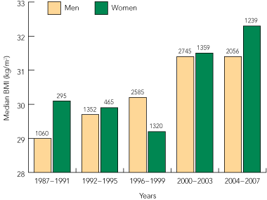
Over the study period, median body weight increased from 89 kg to 99 kg (11%) for men and from 73 kg to 85 kg (16%) for women. This equates to an increase of 0.61 kg/year for men and 0.52 kg/year for women and corresponds with a yearly increase in median BMI of 0.15 kg/m2 for men and 0.14 kg/m2 for women (Box 1). The proportion of individuals with morbid obesity rose more than fivefold, from 3% of the total in 1987 to 16% in 2007.
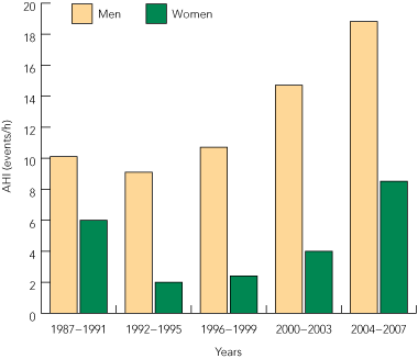
The median AHI for all subjects decreased from 8.8 events/h in the 1987–1991 period to 6.5 in 1992–1995 (P < 0.001), and then rose progressively after that. In 2004–2007, the median AHI was 14.3 events/h. Median AHI was consistently higher in men; however, its rate of increase from 1992 to 2007 was more marked in women (Box 2). The median AHI over this period increased by 107% in men (from 9.1 to 18.8 events/h), but by 325% in women (from 2.0 to 8.5 events/h). For 1992–2007, we calculated an annual rate of increase in the number of subjects with AHI > 30 events/h of just under 1%.
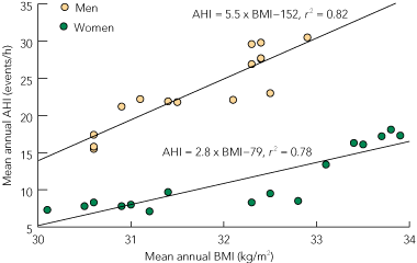
There was a strong correlation between mean annual AHI and mean annual BMI for the period 1992–2007 — for every unit increase in BMI, there was an increase in AHI of 5.5 events/h for men and 2.8 events/h for women (Box 3). Overall, around 80% of the observed variance in AHI was attributable to variance in BMI.
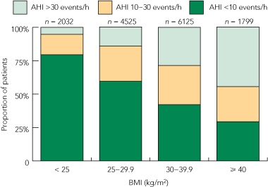
The distribution of severity of SDB according to BMI range is shown in Box 4. This illustrates the relative worsening of SDB severity with increasing obesity: only 5% of individuals with BMI in the not overweight range had an AHI of over 30 events/h, whereas 45% of morbidly obese individuals had an AHI in this range.
Over all the diagnostic studies performed during the 21-year study period, there was a highly significant correlation between AHI and age, with an average increase in AHI of 2.0 events/h for every 10 years’ increase in age (r2 = 0.0149; P < 0.0001).
Our data do not support the hypothesis that increased access to sleep services and increased awareness of sleep disorders is resulting in a decline in disease severity. The finding that about 80% of the observed variance in AHI since 1992 in our study sample is attributable to increases in BMI suggests that the observed worsening in severity of SDB is primarily attributable to increasing obesity.
Published data in this area have not documented long-term trends in severity of SDB. A study from a sleep laboratory in Winnipeg, Canada,11 provides data showing trends in BMI for patients referred for suspected sleep-related breathing disorders over a similar period to our study, but does not provide data on disease severity. During the period 1995–2004, increases in BMI of 3.4 kg/m2 for women and 1.7 kg/m2 for men were reported. Our data over the same period showed increases of around two-thirds of these values (2.4 kg/m2 and 1.2 kg/m2, respectively).
The rate of body weight increase for subjects referred for polysomnography in our study is slightly higher than but similar in magnitude to that documented for the Australian population by the national health survey7 over the period 1989–2005 (0.41 kg/year and 0.34 kg/year for men and women, respectively, compared with 0.61 kg/year and 0.52 kg/year from our data). Projections indicate that rates of overweight and obesity in Australia will continue to increase, with nearly two-thirds of the adult population in these categories by 2019.6 It is therefore not unreasonable to expect that the increases in body weight in those referred for polysomnography will continue, as will corresponding increases in AHI.
We speculate that the higher median AHI observed during 1987–1991 compared with 1992–1995 reflects the selection of subjects with the most severe disease due to the limited number of sleep laboratory beds available at the time. Further, due to the relatively low profile and knowledge of sleep-related disorders at the time, only those with the most clinically overt symptoms and signs of SDB were likely to have been referred for sleep studies. The decrease in AHI in the early 1990s may reflect increased accessibility of services and also a broadening of sleep studies for the investigation of hypersomnolence and fatigue (to which we also attribute the fall in BMI for women during the 1990s). Since the early 1990s, there has been a progressive rise in AHI that is strongly correlated with BMI.
It is tempting to assume that our finding of progressively rising AHI since the early 1990s indicates a progressive increase in the severity of SDB in the general population. However, there is a potential for referral bias as our data represent only subjects referred for diagnostic polysomnography in our laboratory and may reflect the referral patterns of our clinicians rather than be an accurate representation of trends in disease prevalence and severity. On the other hand, ours was the only sleep centre available in the Hunter New England region until 2005, and the only public facility for the duration of the study period. Irrespective of this potential bias, the highly significant correlation between the observed variance in AHI since 1992 and the increase in BMI, coupled with the known increase in obesity rates in Australia over this period, provides solid evidence that severity of SDB is worsening.
The relationship between body-weight gains and SDB severity has been investigated longitudinally in a random population sample of middle-aged adults.8 This analysis revealed, on average, a 32% increase in AHI for a 10% weight gain, corresponding to a mean increase in AHI of 0.15 events/h per kilogram increase in body weight.8 Using our regression data from 1992 onwards, we found a much higher average increase in AHI, of 2.3 events/h per kilogram of weight gain. Therefore, likely changes in SDB severity due to increasing obesity cannot be accurately predicted by applying findings from a random population sample to the clinical population of subjects presenting to the sleep laboratory for diagnostic studies.
It is difficult to accurately quantify the effect on measured AHI of the changes in polysomnographic methods made in the year 2000.10 The average effect is thought to be a small increase in AHI.12 This provides a potential confounding effect on our analysis; however, the continued increase in AHI since these new methods were established, coupled with a strong association between AHI and BMI, suggests that our conclusions remain valid.
It was surprising to find no change over time in the median age of subjects referred for diagnostic sleep studies, but we postulate that this may have resulted from two factors operating in opposing directions. The overall Australian population is ageing, such that median age increased by more than 5 years over the period of this study.13 Secondly, the marked increase in rates of childhood overweight and obesity over the period from 1985 to 1995 has resulted in an increase in young adult obesity.5 This would be expected to have caused a concomitant increase in sleep disorders in this age group that may not have been as evident earlier.
Obese subjects accounted for the majority (78%) of subjects with an AHI of greater than 30, while those with a BMI in the healthy range rarely showed an AHI in this range (5%). Our data reveal that individuals with morbid obesity referred for a diagnostic sleep study had a 45% chance that AHI would be above 30 events/h, whereas there was only a 5% chance for a subject with BMI of less than 25 kg/m2. This type of analysis may be useful in estimation of pre-test probability of significant SDB before formal diagnostic polysomnography.
Our finding of an average increase in AHI of about 2 events/h for every 10 years’ increase in age should be interpreted with caution. It is based on a simple linear regression of cross-sectional data, and, while incidence of sleep apnoea has been shown to increase in older people, it is now recognised that the nature of the relationship between age and sleep apnoea is complex.14 It may not be appropriate to use these data alone to make projections of likely changes in disease severity due to ageing alone.
Overall, our analysis of 21 years of data provides valuable information for planning the future provision of clinical sleep services. Trends indicate that adult sleep services can expect subjects who weigh more, have higher AHIs and represent a more balanced mix of men and women. There are also implications for provision of treatment options based on AHI values. For example, our data indicate that the number of subjects with AHI of over 30 events/h has been increasing at a rate of about 1% per year since 1992. There are obvious cost implications should this criteria be used as the basis for government-funded provision of continuous positive airway pressure therapy. Our data allow projections of the likely costs involved in ongoing management of SDB.
Access to diagnostic sleep laboratory services in NSW changed markedly over the study period; we estimate the number of diagnostic polysomnography beds rose from six in 1987 to over 100 in 2008. The per capita provision of government-funded (Medicare) polysomnography almost trebled (from 123 to 308 studies per 100 000 Medicare-enrolled population) over the decade from 1995 to 2004, indicating increased availability and use of this service.2 The continuing trend of increasing obesity in the Australian population5-7 and the likely implications of this for severity of SDB, coupled with the observation that the per capita provision of sleep studies in Australia was recently shown to be behind that of North America,2 indicates that there will be an ongoing higher demand for access to sleep laboratory services and treatment provision in Australia over coming years. Furthermore, the continued ageing of the Australian population (a projected increase of 3 years in median age by 202615) would be expected to also contribute to worsening incidence and severity of SDB. These issues further highlight the need for more detailed assessment of the roles of non-laboratory-based diagnostic paradigms such as structured screening approaches to diagnosis and home-based investigations.
1 Median body mass index (BMI) of subjects undergoing sleep studies, 1987–2007*
 |
|
* The values above each column indicate the number of sleep studies used in the calculations. |
Received 18 January 2010, accepted 18 April 2010
- Jeffrey J Pretto1,2
- Stephen G Gyulay1,2
- Michael J Hensley1,2
- 1 Department of Respiratory and Sleep Medicine, John Hunter Hospital, Newcastle, NSW.
- 2 School of Medicine and Public Health, University of Newcastle, Newcastle, NSW.
None identified.
- 1. Sullivan CE, Issa FG, Berthon-Jones M, Eves L. Reversal of obstructive sleep apnoea by continuous positive airway pressure applied through the nares. Lancet 1981; 1: 862-865.
- 2. Marshall NS, Wilsmore BR, McEvoy RD, et al. Polysomnography in Australia — trends in provision. J Clin Sleep Med 2007; 3: 281-284.
- 3. Tremblay MS, Katzmarzyk PT, Willms JD. Temporal trends in overweight and obesity in Canada, 1981–1996. Int J Obes Relat Metab Disord 2002; 26: 538-543.
- 4. Young T, Peppard PE, Gottlieb DJ. Epidemiology of obstructive sleep apnea: a population health perspective. Am J Resp Crit Care Med 2002; 165: 1217-1239.
- 5. Venn AJ, Thomson RJ, Schmidt MD, et al. Overweight and obesity from childhood to adulthood: a follow-up of participants in the 1985 Australian Schools Health and Fitness Survey. Med J Aust 2007; 186: 458-460. <MJA full text>
- 6. Sassi F, Devaux M, Cecchini M, Rusticelli E. The obesity epidemic: analysis of past and projected future trends in selected OECD countries. Organisation for Economic Co-operation and Development Health Working Papers, No 45. Paris: OECD Publishing, 2009. doi: 10.1787/225215402672.
- 7. Australian Bureau of Statistics. Overweight and obesity in adults, Australia, 2004–05. Canberra: ABS, 2008. (ABS Cat. No. 4719.0.) http://www.abs.gov.au/ausstats/abs@.nsf/mf/4719.0/ (accessed Aug 2009).
- 8. Peppard PE, Young T, Palta M, et al. Longitudinal study of moderate weight change and sleep-disordered breathing. JAMA 2000; 284: 3015-3021.
- 9. Rechtschaffen A, Kales A, editors. A manual of standardized terminology and scoring system for sleep stages of human subjects. Los Angeles: Brain Information Service, Brain Research Institute, University of California, 1968.
- 10. Sleep-related breathing disorders in adults: recommendations for syndrome definition and measurement techniques in clinical research. The Report of an American Academy of Sleep Medicine Task Force. Sleep 1999; 22: 667-689.
- 11. Banno K, Walld R, Kryger MH. Increasing obesity trends in patients with sleep-disordered breathing referred to a sleep disorders center. J Clin Sleep Med 2005; 1: 364-366.
- 12. Manser RL, Rochford P, Pierce RJ, et al. Impact of different criteria for defining hypopneas in the apnea-hypopnea index. Chest 2001; 120: 909-914.
- 13. Australian Bureau of Statistics. Population by age and sex, Australian states and territories, Jun 2008. Canberra: ABS, 2008. (ABS Cat. No. 3201.0.) http://www.abs.gov.au/AUSSTATS/abs@.nsf/allprimarymainfeatures/2DB211BA9B6E1A25CA2576860017C2F8?opendocument (accessed Aug 2009).
- 14. Young T, Peppard PE, Gottlieb DJ. Epidemiology of obstructive sleep apnoea. A population health perspective. Am J Respir Crit Care Med 2002; 165: 1217-1239.
- 15. Australian Bureau of Statistics. Population projections, Australia, 2006–2101. Canberra: ABS, 2008. (ABS Cat. No. 3222.0.) http://www.ausstats.abs.gov.au/ausstats/subscriber.nsf/0/0E09CCC14E4C94F6CA2574B9001626FE/$File/32220_2006%20to%202101.pdf (accessed Aug 2009).
Abstract
Objective: To document trends in subject demographics, anthropometry and sleep disorder severity over 21 years of diagnostic sleep studies.
Design, participants and setting: A retrospective observational study of consecutive subjects undergoing initial diagnostic polysomnography for investigation of possible sleep disorders in a university-affiliated tertiary public metropolitan hospital in the Hunter New England region of New South Wales between 1987 and 2007.
Main outcome measures: Body weight, body mass index (BMI) and severity of sleep-related breathing disorders (apnoea-hypopnoea index [AHI]).
Results: Between 1987 and 2007, 14 648 new diagnostic sleep studies were performed. The median age of subjects (51 years; interquartile range, 41–61 years) did not change over time and the proportion of women increased from 20% to 39%. Median body weight increased from 89 kg to 99 kg for men (11%) and from 73 kg to 85 kg for women (16%), equating to a yearly increase in median BMI of 0.15 kg/m2 for men and 0.14 kg/m2 for women. The proportion of subjects who were morbidly obese (BMI ≥ 40) increased from 3% in 1987 to 16% in 2007. Median AHI progressively increased from 1992–1995 to 2004–2007 (from 6.5 events/h to 14.3 events/h; P < 0.001), indicating increasing disease severity. Over the same period, for every unit increase in BMI, AHI increased by 5.5 events/h for men and by 2.8 events/h for women. About 80% of the observed variance in AHI over this period was attributable to variance in BMI.
Conclusion: There is a continuing trend towards increasing body weight and BMI in people undergoing diagnostic sleep studies. Our data do not support the hypothesis that increased accessibility to diagnostic services and increased awareness of sleep disorders are resulting in a decline in disease severity. These findings are consistent with the premise that worsening severity in sleep-disordered breathing is primarily attributable to increasing obesity.