


Online responses are no longer available. Please refer to our instructions for authors page for more information.
Connect

Advertisement
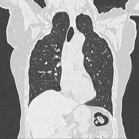
Apical lung hernia
Med J Aust 2007; 187 (6): 368. || doi: 10.5694/j.1326-5377.2007.tb01268.x
Published online: 17 September 2007
Published online: 17 September 2007
Advertisement
Acknowledgement: I thank Dr Bhavin Jankharia, Consultant Radiologist, JIC, Mumbai, India, for the computed tomography image.