Connect
MJA
MJA

Scurvy in an otherwise well young man

Sally J Mapp and Paul B Coughlin
Med J Aust 2006; 185 (6): 331-332. || doi: 10.5694/j.1326-5377.2006.tb00589.x
Published online: 18 September 2006

Clinical record

A 22-year-old male kitchen-hand presented with a 4-week history of extensive bruising and petechiae on his legs (Figure). He had no history of any medical conditions, except for several episodes of epistaxis in the past few months. He was not taking any medication, and there was no known familial bleeding tendency.

On review, he had a large tense haematoma on his left leg that was restricting his mobility, and several smaller bruises in various stages of resolving, along with a palpable petechial rash on both legs (Figure inset). He was unable to recall any trauma that may have precipitated most of the bruises, but, on further questioning, believed that the rash may have been present for up to 2 years. He had no bruising elsewhere. His gums were swollen and bleeding, and oral hygiene was poor. Further examination was unremarkable.

Systematic questioning revealed a long history of a poor diet comprised mainly of carbohydrates, with no vegetable or fruit intake. He smoked 10–15 cigarettes daily and episodically drank large amounts of alcohol. He lived with his girlfriend. A review of other systems showed no abnormalities.

The results of initial investigations, including haemoglobin level, white cell count, and platelet count, were normal, but a blood film showed hypochromic, microcytic red cells. Liver function tests showed no abnormality; and levels of urea, electrolytes and creatinine were normal, as was prothrombin time. Activated partial thromboplastin time was increased at 39 s (reference range [RR], 26–34 s), with full correction on addition of normal plasma.

Further investigations included a repeat measurement of activated partial thromboplastin time (34 s); and a test for levels of factors IX, XI and XII, which gave normal results, whereas the level of factor VIII was raised (249% [RR, 50%–150%]). A screen for lupus anticoagulant was negative, and assays for von Willebrand factor antigen and ristocetin cofactor gave normal results. The results of iron studies showed: iron, 6 μmol/L (RR, 9–27 μmol/L); transferrin, 3 g/L (RR, 2–3.6 g/L); transferrin saturation, 9% (RR, 20%–50%); and ferritin, 83 μg/L (RR, 50–150 μg/L). Finally, the level of vitamin B12 was within the normal range, serum folate level was > 45 nmol/L (RR, 12–32.7 nmol/L); and complement assays (C3, C4) gave normal results. On review 2 weeks later, a repeat blood test showed a low haemoglobin level at 72 g/L (RR, 130–185 g/L). There was no history of altered bowel habit or melaena, and the patient refused a rectal examination. Haptoglobin and lactate dehydrogenase levels were normal.

Because of his iron deficiency and the possibility of significant occult blood loss, he was given a transfusion of packed red cells. The haemoglobin level remained stable after the transfusion.

Further testing revealed a low level of vitamin C (3 μmol/L [RR, 26–85 μmol/L]), and he was prescribed oral vitamin C and iron supplements. Within a week, the condition of his legs was starting to improve. On review 1 month later, their appearance was almost back to normal.

Patient’s left leg at presentation: extensive bruising and petechiae. Inset: perifollicular haemorrhages.

Humans are one of the few mammals that cannot synthesise vitamin C (ascorbic acid). Stores are readily depleted, with studies showing clinical manifestations after about a month on a vitamin C-free diet.1 Vitamin C is absorbed by an energy-dependent, saturable transport system throughout the length of the small intestine, with any excess excreted by the kidneys. Ascorbic acid promotes the hydroxylation of proline and lysine residues in procollagen, helping to stabilise the collagen triple helix. It is the defect in collagen synthesis that produces most of the clinical manifestations.

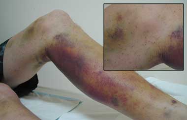
Vitamin C is particularly abundant in citrus fruit and many green vegetables. Scurvy is usually seen in mentally ill patients, people on “fad” diets, people who misuse alcohol, and, occasionally, elderly patients who live alone.2 Initial symptoms are non-specific, including weakness, anorexia, depression and lethargy. Subsequent dermatological features include broken and coiled hairs (due to abnormal keratin formation) and perifollicular haemorrhages and hyperkeratosis. Bleeding tends to be a late feature and affects spongy, friable gums as well as muscles, joints and skin. This is thought to be due to breakdown of the connective tissue within and supporting the vessel walls, rather than a platelet or clotting factor defect.3 Many patients with scurvy are anaemic, with contributing causes likely to be bleeding, altered absorption and metabolism of iron and folate,3 and other dietary deficiencies.

Our patient is interesting in that he does not seem to fit into the usual groups with a tendency to develop vitamin C deficiency. A full dietary history revealed a diet entirely comprised of Vegemite spread, cheese, bread, dry biscuits, chocolate and a cola drink, with no fruit or vegetables. The patient conceded that he had not tried any new foods for over 10 years, and, despite the recent diagnosis, was reluctant to initiate dietary change.

Although such a limited food intake is probably uncommon, recent trends towards consumption of pre-prepared carbohydrate-rich food mean that scurvy or subclinical vitamin C deficiency and other vitamin and trace metal deficiencies should not be forgotten.

  • Sally J Mapp1
  • Paul B Coughlin2

  • 1 Department of Haematology, The Canberra Hospital, Canberra, ACT.
  • 2 Australian Centre for Blood Diseases, Melbourne, VIC.


Correspondence: smapp@member.rcpa.edu.au

Competing interests:

None identified.

  • 1. Padayatty SJ, Levine M. New insights into the physiology and pharmacology of vitamin C. CMAJ 2001; 164: 353-355.
  • 2. Case records of the Massachusetts General Hospital. Weekly clinicopathological exercises. Case 39-1995. A 72-year-old man with exertional dyspnea, fatigue, and extensive ecchymoses and purpuric lesions. N Engl J Med 1995; 333: 1695-1702.
  • 3. Reuler JB, Broudy VC, Cooney TG. Adult scurvy. JAMA 1985; 253: 805-807.

Author

remove_circle_outline Delete Author
add_circle_outline Add Author

Comment
Do you have any competing interests to declare? *

I/we agree to assign copyright to the Medical Journal of Australia and agree to the Conditions of publication *
I/we agree to the Terms of use of the Medical Journal of Australia *
Email me when people comment on this article

Online responses are no longer available. Please refer to our instructions for authors page for more information.