


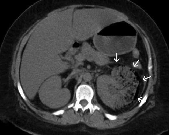
A 40-year-old Indigenous woman with type 2 diabetes presented with a 6-month history of intermittent left flank pain. She reported worsening pain of 2 days’ duration associated with fever, nausea, vomiting and reduced urine output. Clinical examination revealed tenderness over the left loin. An x-ray (not shown) and computed tomogram of the abdomen (Box) suggested a diagnosis of emphysematous pyelonephritis.
Emphysematous pyelonephritis is a rare, severe gas-forming infection of the renal parenchyma, typically seen in people with diabetes. Radiologically, four classes of emphysematous pyelonephritis are described on computed tomography:1 in Class 1 and 2, the gas is localised to the collecting system and the renal parenchyma, respectively, without extension to the extrarenal space; in Class 3A, as seen in this case, there is extension of gas or abscess into the perinephric space, and in Class 3B, to the pararenal space; bilateral emphysematous pyelonephritis or emphysematous pyelonephritis of a solitary kidney represents the most severe form of the disease (Class 4).
Emphysematous pyelonephritis is associated with a high mortality rate (40%) when treated with antibiotics alone.1 Although milder forms of the disease (Class 1 and 2) have been successfully treated with a combination of percutaneous drainage and antibiotics, these modalities alone may not be sufficient in more severe presentations of the disease or in patients presenting with septic shock. In such patients, early nephrectomy is recommended.1,2
- 1. Huang JJ, Tseng CC. Emphysematous pyelonephritis: clinicoradiological classification, management, prognosis, and pathogenesis. Arch Intern Med 2000; 160: 797-805.
- 2. Adbul-Halim H, Kehinde EO, Abdeen S, et al. Severe emphysematous pyelonephritis in diabetic patients: diagnosis and aspects of surgical management. Urol Int 2005; 75: 123-128.
None identified.