


Although many fractures in children can be managed with pain relief and splinting alone, a thorough knowledge of which ones require referral and urgent intervention is essential
Falls are the most common cause of injury in children presenting to an emergency department.1 Children, with their relatively large heads, plastic bones, and limited ability to protect themselves during a fall, frequently sustain fractures and minor head injuries. Management of the other types of minor childhood injuries that are frequently encountered — bruising, abrasions, and lacerations — is covered in the previous article in the MJA Practice Essentials Series – Paediatrics (Bruising, abrasions, lacerations: minor injuries in children I. Med J Aust 2005; 182: 588-592); fractures and minor head injuries will be dealt with in this article.
As explained in the first article on childhood injuries, we define a minor injury as one that could reasonably be expected to heal with minimal medical intervention. The importance in both fractures and head injuries of distinguishing between “minor” and “major” injuries is emphasised.
In the previous article, we described techniques for increasing the chance of a successful assessment of an injured child, such as gaining the child’s trust, and dealing with pain at an early stage with analgesics, splinting and distraction. Information about analgesia, sedation and local anaesthesia, as well as antibiotics and tetanus prophylaxis, is given in the previous article (see MJA 2005; 182: 588-592).
During assessment of an injured child, the child’s immunisation status, allergies to antibiotics or dressings, and any pre-existing medical conditions need to be established.
Common fractures in children involve the clavicle, humerus, radius, ulna, distal tibia, and fibula. Incomplete fractures involving only one cortex are relatively stable and generally can be managed with splinting to relieve pain and prevent further injury.
It is important to be able to describe fractures accurately for documentation or discussion with a colleague, especially by telephone. Include in the description:
Anatomical site of the fracture — “which bone?”, “which side?”, and “is the fracture in the proximal, middle or distal third of the bone?”;
Appearance of the fracture line — transverse, oblique, spiral, or comminuted (more than one fragment);
Whether the fracture is open or closed;
Angulation and displacement of the fracture:
to determine angulation, draw a line along the edge of both major fragments and measure the angle of intersection of the distal fragment.
to determine the displacement, compare it with the width of the bone at that point (eg, half a diameter displaced or completely displaced).
In metaphyseal fractures (ie, fractures in the expanded ends of long bones), it is important to note the proximity of the fracture to the growth plate, as this determines the remodelling potential of the bone and cartilage around the joint. Injuries to the growth plate are described according to the Salter–Harris classification (Box 1).2
A simple undisplaced buckle fracture with only one cortex involved can be treated with an easily removable plaster or fibreglass slab.3 However, if the fracture involves both cortices of the bone, an encircling plaster or a plaster/fibreglass slab extending across the joint above and below should be used.
If treating a child with an encircling plaster as an outpatient:
Try to get the child to elevate the limb above chest level for 24–48 hours after fitting the plaster. This may not be possible in small children.
A sling should only be worn after this period, and the hand should not be below the elbow while in the sling.
For leg plasters, crutches should only be used by children over 6–7 years of age who have good coordination.
A review of the plaster should be organised for the day after fitting the plaster to check whether it is too tight. Look for pain on passive extension of the fingers or toes, swelling and colour of the digits, and assess distal circulation. Pain at the fracture site alone is rarely a reason to split a plaster.
Go through written instructions explaining the care of a child with a plaster to the parents, and give them a copy. For an example, see <www.rch.org.au/kidsinfo/factsheets.cfm?doc_id=4083>.
For children who have had a fracture manipulated or whose fracture involved both cortices of the bone, a repeat x-ray should be obtained after 1 week to ensure the correct position is maintained. If a plaster slab has been used, then this should be replaced by an encircling plaster, and a repeat x-ray through the plaster obtained. The plaster cast should remain in place for 3–6 weeks, depending on the site and severity of injury. Following the removal of the cast, the bone is still at risk of re-fracture until the fracture consolidates, which may take 6–12 weeks. Contact sports are not recommended during this period.
Management of the various types of upper- and lower-limb fractures, including those requiring urgent referral, are summarised in Box 2 (upper limb), Box 3 (hand), and Box 4 (lower limb).
An ankle sprain can be difficult to distinguish, clinically or radiologically, from a fracture through the growth plate of the distal fibula. Young children with open growth plates are more likely to sustain a growth plate injury or a fracture than a ligament injury and should be treated in a plaster cast for 2 weeks and re-examined. If the site is still painful or a repeat x-ray shows a fracture, maintain immobilisation for another 4 weeks. True sprains are more common in adolescents.
X-rays are required if any of the following are present:
Pain over the posterior edge of the distal tibia or fibula.
Pain over the navicular or head of the fifth metatarsal.
The child is unable to bear weight for four steps both immediately after the injury and later in the surgery.5
These indications for an x-ray examination are commonly referred to as the Ottawa ankle rules.6 Although they are a useful guide, the Ottawa ankle rules may not be as sensitive at predicting a fracture in children as they have proven to be in adults.7
If a child has tenderness over the growth plate of the distal tibia or fibula, and x-rays show no abnormality, treat as a Salter–Harris I epiphyseal injury. If swelling is not marked, apply a below-knee plaster cast for 4–6 weeks. If there is marked swelling, treat in a backslab for 2 weeks, then re-examine. If the area over the growth plate is now no longer tender, mobilise with the assistance of physiotherapy; otherwise, continue the immoblisation. If a fracture is present, an x-ray at this time should show new bone formation.
Treat in a similar way to a fracture: use a below-knee plaster cast (or, if the swelling is severe, a backslab followed by a full plaster cast) for a maximum of 1–2 weeks. No weight-bearing is allowed for the first few days.
Gradual rehabilitation should occur after this.
Physiotherapy is useful in all grades of ankle ligament injury. Functional treatment — brace or tapes, with active physiotherapy — has been shown in older adolescents to produce a better outcome than immobilisation, with greater joint stability and an earlier return to sport.8
A pulled elbow is a common injury in toddlers, but it may occur in children up to the age of 5 years. It is most often caused by a person forcibly pulling on the child’s arm, but it may also result from a fall, or the mechanism may be unknown.9
The most common scenario in head injuries is for a child to fall and hit his or her head, to cry immediately after the fall and become inconsolable for minutes to hours. Younger children may then fall asleep.
A child with no loss of consciousness after the incident and only one or no episodes of vomiting, whose condition is currently stable, and who is alert and interactive, and neurologically normal, is extremely unlikely to have sustained any intracranial injury. Such a child may be safely managed at home. Parents need to have clear instructions, especially about when to seek urgent medical advice. This should occur if their child:
becomes unconscious or difficult to rouse;
becomes confused;
has a fit;
develops a persistent headache;
repeatedly vomits; or
develops any bleeding or watery discharge from the ears or nose.
Instructions for parents of a child who has sustained a minor head injury are available at: <http://www.rch.org.au/kidsinfo/factsheets.cfm?doc_id=3728>.
Signs requiring immediate referral to hospital include:
persisting depression, or deterioration, in conscious state;
focal neurological signs;
possible penetrating skull wound; or
persistent headache.
Children developing these signs require an urgent computed tomography scan of the brain and possible neurosurgical intervention.10
Evidence-based practice tip
Fibreglass backslabs or moulded splints are a suitable alternative to traditional full casts in children with a minimally displaced buckle fracture of the distal radius (III).3
Levels of evidence (I–IV) are derived from the National Health and Medical Research Council’s system for assessing evidence.11
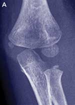
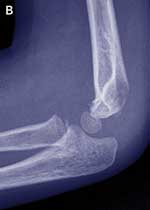
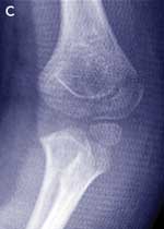
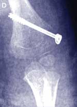
Case study — a 4-year-old girl with a painful left elbow after a fall
The parents of a 4-year-old girl bring their daughter to you after she has fallen about 1 metre from climbing equipment onto her outstretched arm. The girl is complaining of pain in her left elbow. She is unable to straighten her arm without pain and can only flex to 110°. The elbow looks swollen, mainly on the medial aspect. She has diffuse tenderness around the elbow and is unable to supinate or pronate her arm. There is no neurovascular compromise. Management
|
|||||||||||||||
  |
|||||||||||||||
  |
|||||||||||||||
Lateral condyle fractures are inherently unstable and take a longer time to heal than supracondylar fractures. They should be managed by internal fixation in most cases. This case illustrates that it may be difficult to diagnose these fractures and that close follow-up is needed to prevent complications. |
|||||||||||||||
1 Salter–Harris classification of epiphyseal injuries
Type I, fracture through the growth plate; Type II, fracture through the growth plate and metaphysis; Type III, fracture through the growth plate and epiphysis; Type IV, fracture through the growth plate, epiphysis and metaphysis; and Type V, crush or compression injury of the growth plate.
2 Upper limb fractures (excluding hand fractures)
Type of fracture |
Important issues |
When to refer immediately |
Management |
||||||||||||
Clavicle |
A lump will develop at the fracture site which may be visible for 1 year No follow-up x-ray is necessary |
Fracture is tenting the skin Involvement of acromioclavicular joint Brachial plexus injury |
Broad arm sling for 3–4 weeks Adequate analgesia No contact sports for at least 6 weeks No medical follow-up necessary4 |
||||||||||||
Surgical neck of humerus |
|
Displaced or markedly angulated |
Undisplaced — sling for 3 weeks |
||||||||||||
Shaft of humerus |
Check the integrity of the radial nerve |
Transverse, displaced, comminuted |
Undisplaced — collar and cuff, with U-shaped slab to reduce movement and minimise pain. Remove the U slab after 2–3 weeks and leave the arm in a sling until pain resolves |
||||||||||||
Supracondylar region of humerus |
Check the integrity of the radial artery, radial nerve, median nerve and ulnar nerve Swollen elbow with joint effusion only indicates an undisplaced fracture — look for displacement of the anterior and posterior fat pads on x-ray |
Angulated, displaced or comminuted Look carefully for undisplaced lateral condyle fractures — refer if displaced or any doubt about the diagnosis (see Case study) Vascular compromise needs to be corrected within 4 hours of injury |
If vascular compromise is present, extend the elbow until perfusion returns and refer urgently Undisplaced — collar and cuff (under clothing) or backslab with the elbow flexed to 110° for 3–4 weeks |
||||||||||||
Shaft of radius and/or ulna |
Isolated fracture of ulna — look for radial head dislocation (on lateral x-ray) Isolated radial fracture — look for distal radioulnar joint dislocation |
Displaced, angulated, open Displacement of the radial head or distal radioulnar joint |
Undisplaced — above-elbow cast |
||||||||||||
Distal radius with or without ulna |
Single cortex affected — fracture may appear to be angulated when not Angulations of up to 20o may be acceptable — the older the patient the lesser the degree of angulation allowed If arm looks deformed, then reduction of fracture required |
Arm appears deformed Displaced, angulated > 20o — will require manipulation |
If a single cortex only is involved, use a short arm cast or backslab for 2–4 weeks Remove cast when fracture site no longer tender If both cortices involved — above-elbow plaster. If angulation increasing, then urgent referral required |
||||||||||||
3 Hand fractures
Type of fracture |
Important issues |
When to refer immediately |
Management |
||||||||||||
Metacarpals |
Fifth is the commonest Look for full extension of fingers Look for rotation of finger with flexion of finger Acceptable angulation of a metacarpophalangeal neck fracture is 20°, 2nd; 30°, 3rd; 40°, 4th; 50°, 5th |
Angulated or rotated shaft fracture (will require pinning) |
Undisplaced or acceptable angulation — volar slab with hand flexed at the distal interphalangeal/metacarpophalangeal joint for 3 weeks |
||||||||||||
Phalanges |
Check carefully for rotation at the fracture site (particularly, distal end of proximal phalanx) Intra-articular fractures require anatomical reduction |
Displaced or angulated |
Undisplaced — strap to the adjacent finger for 3–4 weeks |
||||||||||||
Mallet finger |
Inability to extend distal phalanx |
Large bone fragment associated with injury |
Mallet splint for 6 weeks non-stop (ie, not to be removed, even to shower) |
||||||||||||
4 Lower limb fractures
Type of fracture |
Important issues |
When to refer immediately |
Management |
||||||||||||
Tibial fracture |
Should be suspected in a child not bearing weight after an injury |
Displaced, angulated |
Proximal or midshaft — above-knee plaster Distal — below-knee plaster |
||||||||||||
“Toddlers fracture” (undisplaced tibial or fibula fracture not seen on initial x-ray) |
Clinical diagnosis in a young child not bearing weight after an injury and/or with tenderness on palpation of tibia/fibula; x-ray looks normal |
|
Allow as much weight-bearing as the child desires if the pain is minimal, or, if pain at rest or nocturnal pain, fit a below-knee plaster for 2–3 weeks Limping will continue for 6 weeks |
||||||||||||
Distal tibia/fibula |
Refer if articular surface of tibia involved |
Displaced, angulated, involving both tibia and fibula |
Undisplaced — below-knee plaster for 6 weeks |
||||||||||||
Metatarsals |
|
|
Undisplaced or displaced — lower leg plaster slab for 4 weeks. Elevation for the first few days will help minimise swelling |
||||||||||||
- Simon J Young1
- Peter L J Barnett2
- Ed A Oakley3
- Department of Emergency Medicine, Royal Children’s Hospital, Parkville, VIC.
This article is based on the clinical practice guidelines of the Royal Children’s Hospital, Melbourne. These are readily available on the Internet at
- 1. Clapperton A, Ashby K, Cassell E. Injury profile Victoria 2001. Hazard 2001; 54: 1-24. Available at: www.monash.edu.au/muarc/VISAR/hazard/haz54.pdf (accessed May 2005).
- 2. Salter RB, Harris WR. Injuries involving the epiphyseal plate. J Bone Joint Surg Am 1963; 45: 587-622.
- 3. Howes M. Bracing for buckle fractures of the distal radius in children. Available at: www.bestbets.org/cgi-bin/bets.pl?record=00349 (accessed Mar 2005).
- 4. Calder JD, Solan M, Gidwani S, et al. Management of paediatric clavicle fractures — is follow-up necessary? An audit of 346 cases. Ann R Coll Surg Engl 2002; 84: 331-333.
- 5. Plint AC, Bulloch B, Osmond MH, et al. Validation of the Ottawa ankle rules in children with ankle injuries. Acad Emerg Med 1999; 6: 1005-1009.
- 6. Stiell IG, Greenberg GH, McKnight RD, et al. Decision rules for the use of radiography in acute ankle injuries. JAMA 1993; 269: 1127-1132.
- 7. Clark KD, Tanner S. Evaluation of the Ottawa ankle rules in children. Pediatr Emerg Care 2003; 19: 73-78.
- 8. Kerkhoffs GM, Rowe BH, Assendelft WJ, et al. Immobilisation and functional treatment for acute lateral ankle ligament injuries in adults. Cochrane Database Syst Rev 2002 (3): CD003762.
- 9. Schutzman SA, Teach S. Upper extremity impairment in young children. Ann Emerg Med 1995; 26: 474-479.
- 10. Munro A, Maconochie I. Indications for head CT in children with mild head injury. Emerg Med J 2001; 18: 469-470.
- 11. National Health and Medical Research Council. How to use the evidence: assessment and application of scientific evidence. Handbook series on preparing clinical practice guidelines. Table 1.3: Designation of levels of evidence. Canberra: NHMRC, February 2000: 8. Available at: www.health.gov.au/nhmrc/publications/pdf/cp69.pdf (accessed Mar 2005).
Abstract
Fractures in children are common, but the plasticity of children’s bones means that they may be incomplete.
If a child has deformity, swelling or bony point tenderness in a limb after a fall, it is likely to be fractured.
A fractured limb that appears deformed will most probably need to be reduced.
Effective splinting, using whatever means is readily available, and early, adequate analgesia, can ameliorate the severe pain associated with a fracture.
In young children with open growth plates, Salter–Harris type I injuries of the distal fibula are more common than ligament injuries of the ankle.
After an ankle ligament injury, functional treatment — brace or tapes, with active physiotherapy — results in a better outcome than immobilisation.
A child with a head injury, who does not lose consciousness, has only one or no episodes of vomiting, and is stable, alert and interactive, and neurologically normal, is extremely unlikely to have sustained an intracranial injury.